Jeffrey Landis [not his real name], 74, had been complaining of swallowing problems for a couple of months. His wife urged him to go to the doctor, but he delayed a visit, thinking that his symptoms would resolve. But one day, during his afternoon duties as a reading tutor, he regurgitated a piece of sandwich he had eaten at lunch-and it was undigested. In addition to his embarrassment, Mr. Landis was alarmed, and called up his otolaryngologist’s office for a visit the next day.
Explore This Issue
April 2009Otolaryngologists-head and neck surgeons would most likely identify Mr. Landis’s symptoms as suspicious for esophageal diverticulum. How they would go about establishing a definitive diagnosis and planning treatment was a topic ENT Today explored recently with head and neck surgeons who see patients with esophageal diverticula in their practices.
Prevalence and Presentation
Zenker’s diverticulum (ZD) is the most common type of esophageal diverticula, and develops in the triangular space between the inferior pharyngeal constrictor muscle and the cricopharyngeus muscle. Although its etiology has not been definitively established, ZD appears to be caused by the discoordination between the two muscle groups. Increased pressure in the oropharynx during swallowing against a closed upper esophageal sphincter can cause the hypopharyngeal mucosa to pouch out and prolapse through the triangular space. Food gets diverted into and trapped inside the diverticular pouch instead of passing through the hypopharynx into the esophagus.1,2 (There are other inherent areas of weakness along the esophageal wall, where lateral or Killian-Jamieson diverticula can also occur.)
ZD occurs most often in men in their 70s and 80s, although cases in patients in their 50s are not uncommon. Gregory Postma, MD, Director of the Center for Voice and Swallowing Disorders at the Medical College of Georgia in Augusta, has performed surgical repair on patients as young as 35 years old. The prevalence in the general population, according to a 1996 study, is between 0.01% and 0.11%.3
The true incidence of ZD is difficult to determine, however, as some people with esophageal diverticula are asymptomatic. The classic symptoms are dysphagia and regurgitation. Sometimes there is a bubbling sound coming from the pouch during eating. As the pouch grows bigger, more food gets trapped, Dr. Postma explained, and patients may lose weight due to malnutrition. These patients have trouble eating solids, and often their spouses will report that it takes them a long time to eat their meals, he said.
The condition can also lead to aspiration pneumonia. Symptoms can be present for several years before patients seek treatment.
The Gold Standard
Although endoscopic treatment of esophageal diverticula is now becoming standard of care (see sidebar), endoscopic-based diagnosis is not the usual strategy. The gold standard remains the barium swallow, asserted Dr. Postma.
Alexander T. Hillel, MD, a resident in the Department of Otolaryngology-Head and Neck Surgery at Johns Hopkins School of Medicine in Baltimore, who recently co-authored a historical review of endoscopic surgical management of ZD with Department Chair Paul W. Flint, MD, concurred with this approach: At our institution, when we are referred patients who are demonstrating symptoms of dysphagia and in whom a suspicion for esophageal diverticulum is high, we typically order a barium swallow.
But in the offices of Dinesh K. Chhetri, MD, Assistant Professor of Head and Neck Surgery and Director of the Swallowing Disorders Center at the David Geffen School of Medicine at the University of California, Los Angeles, patients with symptoms such as Mr. Landis’s might just as likely undergo fiberoptic endoscopic evaluation of swallowing (FEES) followed by transnasal esophagoscopy (TNE) to diagnose their problem.
During the January 2009 meeting of the Triological Society’s Western Section, Dr. Chhetri and his research assistant Jennifer Long, MD, PhD, presented results of a retrospective cohort review, in which they reported that the finding of esophagopharyngeal reflux (EPR) when performing FEES has a high sensitivity and specificity for the presence of an esophageal diverticulum. TNE is then used to visualize and confirm the diagnosis of diverticulum during the same office visit, and the patient can be scheduled for surgery. I don’t find that the barium swallow is necessary anymore if I can diagnose the Zenker’s diverticulum in my office, Dr. Chhetri said recently from his Los Angeles office. With the knowledge that the pouch is there, I can then go ahead and schedule the patient for surgery.
Dr. Postma, who has published widely on the applications of FEES and TNE, said, When I do FEES and find an abnormality, very often it leads us to another diagnostic test. So, for example, if regurgitation occurs, that person is going to get a barium swallow. Many other conditions can account for the presence of EPR, he noted-including proximal esophageal strictures, severe esophageal dismotility, and tumors. Some small ZDs do not demonstrate EPR, and therefore would be missed if a barium swallow was not obtained. In addition, added Dr. Postma, obtaining a barium swallow allows me to look at the vertebral bodies for the presence of osteophytes. Large osteophytes sometimes make endoscopic exposure of a ZD more difficult.
The FEES/TNE Diagnostic Process
Dr. Chhetri maintained that his methods for diagnosing diverticula offer a way to overcome what he calls the shortcomings of traditional radiologic evaluation-that is, diagnosis and treatment planning do not have to be delayed. Because many otolaryngologists now routinely perform FEES and TNE, they can more readily diagnose and plan treatment in one patient visit. (This may be more of an issue in private practice than at an academic center, where radiology is generally housed in the same building or on the same campus.)
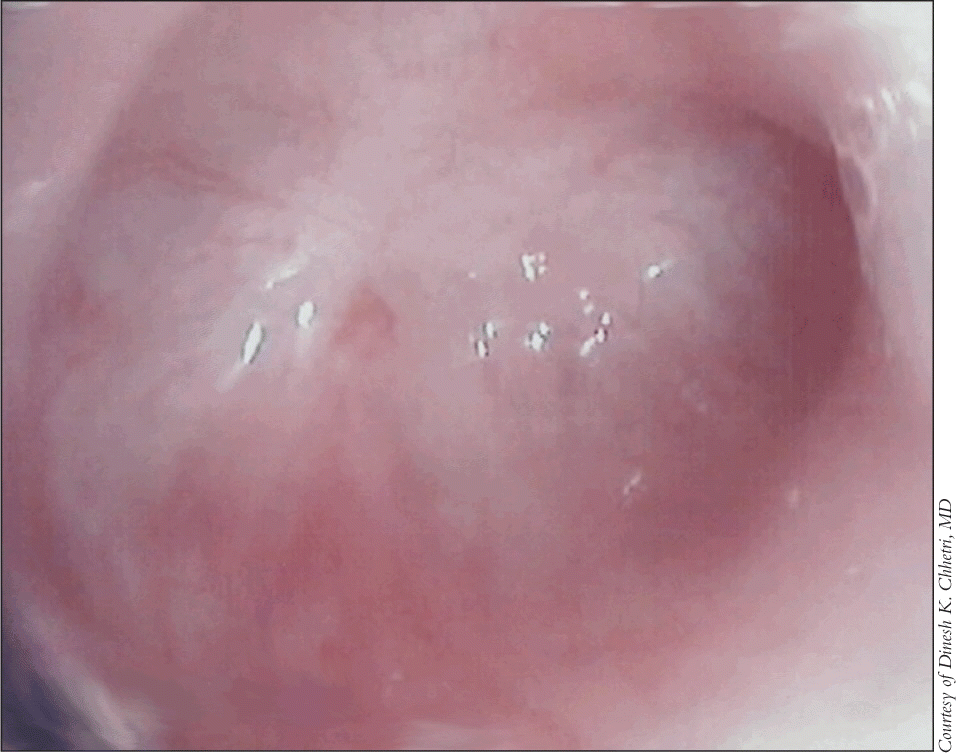
When performing FEES, Dr. Chhetri first uses a 4% neosynephrine solution, without topical anesthetic, to decongest the nasal passages. After the scope has been passed along the floor of the nose and into the oropharynx, food mixed with green coloring is fed to the patient, beginning with puree, followed by nectar-thick, thin liquids, and a cookie. If patients are suspected to have a diverticulum, the endoscopist performs additional maneuvers to provoke EPR. These include asking the patient to phonate a sustained vowel (eeeee) after swallowing the food bolus and/or pressing on or massaging the patient’s anterior neck during sustained phonation. In his experience, said Dr. Chhetri, these maneuvers can cause the contents of a cervical esophageal diverticulum to be refluxed back to the hypopharynx. Observance of EPR would then lead Dr. Chhetri to perform TNE. After anesthetizing the nasal cavity with 4% lidocaine-soaked pledgets for several minutes, the transnasal esophagoscope is passed through the nasal cavity, advanced to the hypopharynx, and directed into a pyriform sinus, then allowed to pass into the cervical esophagus during a swallow.
If a ZD is present, the tip falls naturally into the diverticular pouch. Dr. Chhetri contended that he can generally estimate the pouch size by noting the distance the endoscope travels from the tip of the esophageal pouch to the level of the cricopharyngeal bar. After assessing the diverticulum, the scope is withdrawn to the cricopharyngeal bar and redirected toward the esophagus. The entire esophagus is then assessed all the way to the stomach.
Hindrance to Surgical Planning?
Dr. Hillel said that he recommends the standard barium swallow for definitive diagnosis and ease of surgical planning. So does Dr. Postma. The barium swallow gives you the ability to tell the size of the pouch-and size is pretty important, noted Dr. Postma. If the pouch is tiny, he explains, it may be impossible to insert the staple device used to perform the diverticulostomy. In the latter case, a laser myotomy is then performed.
Dr. Chhetri said that knowing the exact size of the diverticulum before he heads into surgery is not necessary. My approach is that if the diverticulum is large enough that I can staple, then I staple it, he said. If it’s small and the stapler doesn’t fit, I use the carbon dioxide laser. If one is prepared for both possibilities, then the technique used to treat the diverticulum can be chosen accordingly in the operating room.
Dr. Postma prefers to prepare his patients before surgery regarding the approach he will take, and the barium swallow allows him to do this with 97% certainty, he said. A patient with a ZD large enough to receive the stapling approach will be fed soon after surgery and sent home either the same day or the next morning, whereas a patient receiving the laser myotomy will not receive food until the day after. There could be other concerns related to this diagnostic and treatment approach, noted Dr. Postma. For instance, the barium swallow is less costly than TNE; obtaining a barium swallow could be wise from a medicolegal standpoint should there be a misadventure during surgery; and certain payers may not reimburse for both endoscopic procedures performed on the same day for the same diagnosis.
Dr. Chhetri added that he doesn’t advocate deleting use of the barium swallow altogether. If you see someone who has dysphagia and you’re not sure of the etiology based on FEES and TNE, then you can always obtain a barium esophagogram or a modified barium swallow study, he said. In his experience, diagnosis of ZD is pretty straightforward using his techniques. Dr. Chhetri estimated that in the past year, he diagnosed and treated approximately one patient per month with Zenker’s. It’s not an everyday thing, but it is a highly treatable condition.
Endoscopic Approach Now Accepted in Surgical Management
According to an historical review of the evolution of endoscopic surgical management for Zenker’s diverticulum written by Alexander T. Hillel, MD, and Paul W. Flint, MD,(2) traditional surgical approaches including the two-stage open diverticulectomy were utilized into the 1950s. By the late 1950s and early 1960s, surgeons began to realize that the diverticulectomy required a myotomy to address the discoordination of the inferior pharyngeal constrictor and spastic cricopharyngeus muscles.
Gösta Dohlman, MD, pioneered an endoscopic esophagodiverticulostomy and myotomy using electrocautery. Then, in the mid-1990s, the endoscopic stapler-assisted diverticulostomy was introduced, simultaneously dividing and stapling the mucosal edges of the esophagodiverticular wall. The stapler alleviated concern with sutureless division of electrocautery and laser techniques. Collard published the first studies validating this approach in 1993.4
Endoscopic stapler-assisted diverticulostomy now represents the first-line surgical treatment for Zenker’s diverticulum because of reduced morbidity and shortened operative and recovery times compared with external approaches, write Drs. Hillel and Flint.
References
- Ferreira LE, Simmons DT, Baron TH. Zenker’s diverticula: pathophysiology, clinical presentation, and flexible endoscopic management. Dis Esophagus 2008; 21:1-8.
- Hillel AT, Flint PW. Evolution of endoscopic surgical therapy for Zenker’s diverticulum. Laryngoscope 2009;119:39-44.
- Watemberg S, Landau O, Avrahami R. Zenker’s diverticulum: reappraisal. Am J Gastroenterol 1996;91:1494-8.
- Collard JM, Otte JB, Kestens PJ. Endoscopic stapling technique of esophagodiverticulostomy for Zenker’s diverticulum. Ann Thorac Surg 1993;56:573-6.
©2009 The Triological Society

Leave a Reply