Explore This Issue
June 2011CHICAGO — A 29-year-old banker with a mild upper respiratory infection that’s been lingering for a week arrives at the otolaryngologist’s office with tinnitus. He had flown six days earlier and lifted at the gym five days earlier.
It’s found that his speech discrimination score is “very poor.” Neither the flight nor his workout seem to have a cause-and-effect relationship with any specific event he’s suffered.
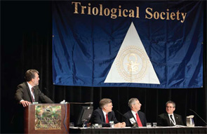
This case was one of three discussed by a panel moderated by Samuel Selesnick, MD, FACS, professor and vice chair of otolaryngology at Weill Cornell Medical College in New York City, on April 29 at the Annual Meeting of the Triological Society, held here as part of the Combined Otolaryngology Spring Meetings.
The panel also discussed the case of a young boy with recurrent cholesteatoma and a classic case of Ménière’s disease.
Tinnitus Troubles
In the case of the banker with tinnitus, the panel agreed there was no obvious answer.
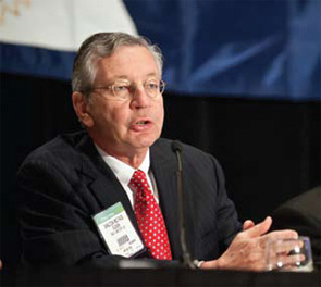
“He certainly has a severe profound left-sided sensorineural hearing loss with poor discrimination,” said D. Bradley Welling, MD, PhD, chair and professor of otolaryngology-head and neck surgery at the Ohio State University Medical Center in Columbus. “And the differential diagnosis for an idiopathic sensorineural hearing loss is quite broad, but you would probably want to evaluate that more.”
Clough Shelton, MD, FACS, professor and chief of otolaryngology-head and neck surgery at University of Utah Health Care in Salt Lake City, agreed that the spectrum starts out wide in a case like this.
“I usually explain this to patients as either being a virus or a vascular event,” he said. “When you look histologically, though,… it’s really a mixed bag of things. I don’t know that we have one etiology that causes this problem.”
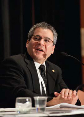
Alan Micco, MD, associate professor of otolaryngology-head and neck surgery at the Northwestern University Feinberg School of Medicine in Chicago, brought up the possibility of a perilymphatic fistula.
“Obviously, he’s not having significant vertigo, but it’s something we have to think about as well in this situation,” he said.
Dr. Shelton played down that possibility. “It certainly suggests the concern of perilymph fistula,” he said. “I’m still a skeptic, though, of perilymph fistulas, because I think that if these types of activities that a patient underwent would cause a fistula, and cause this degree of hearing loss, we’d all have fistulas. We all do this sort of stuff day in and day out. I have less of a level of suspicion in these types of cases for a fistula. I don’t explore very many patients for fistulas these days.”
Dr. Welling agreed that his level of suspicion of a perilymphatic fistula would be “fairly low.”
There was general agreement that it would be worthwhile to do an MRI in this case.

“An MRI is reasonable since a percentage of patients with acoustic tumors present with sudden hearing loss,” Dr. Welling said. “I don’t typically do the full battery of OAE (otoacoustic emissions) and BAERs (brainstem auditory evoked response) unless there’s some other indication. But it seems like, with a good audiogram and ruling out retrocochlear disease with an MRI, that would be useful.”
He said he also might order labs, such as viral titers, if something specific in the history suggested the need, but added that “I don’t always do them on sudden hearing loss.”
Dr. Micco said he doesn’t consider lab tests unless the hearing loss is bilateral. “I think there’s enough evidence, though, especially in patients that have had recovery after sudden loss, there still could be a retrocochlear lesion. So I think an MRI is essentially imperative,” he said.
The panelists also agreed that oral steroids would be the best course. And Dr. Shelton said he often uses a vasodilator in a case like this.
“I usually will treat patients or follow patients for four months and feel like that’s the time period where if they’re going to recover they will recover,” he said. “And so many times after the course of steroids, if they don’t get better, I’ll put them on a vasodilator. I don’t have good evidence, scientific evidence, that it works, but I think that it certainly doesn’t hurt. And it may help some people recover. And [it’s] something that I can treat them with for the full four-month period that I’m watching them.”
Dr. Welling noted that studies have shown that antivirals don’t show any advantage over steroids, but Dr. Micco said he might use them in some cases.
“I’ll have patients that will come in with a sudden (hearing) loss that will have these pain symptoms and some of them do seem to respond to the antivirals,” he said.
Panelists generally agreed that intratympanic therapy is worthwhile as a salvage treatment.
Dr. Selesnick asked, “Is our best argument that it probably won’t hurt you and it may help? In other words, the downside is down? Or is our best argument that there’s a benefit to this? And this is a question that is in evolution.”
Dr. Welling said there was not a clear answer to that. “I want to know the answer to this question myself,” he said. “I have to say I don’t have a good regime yet. We will try it. Most of the time when we see these folks, they’ve already been put on oral steroids by either their primary care physician or by another otolaryngologist in the community. So when they come in and they’re on oral steroids and they haven’t responded, then we’ll certainly offer them transtympanic dexamethasone.”
Dr. Micco said that, like Dr. Welling, he offers patients both oral steroids and intratympanic therapy. “I think that we do miss patients by just giving them intratympanics,” he said. “With that being said,… I offer them both. But I always tell patients we can always salvage and I think, actually, that’s the data that we need. Do we have the ability to salvage these patients in a long-term way?”
Dr. Shelton said he would typically use intratympanic therapy as salvage therapy.
“I’m not a real intratympanic enthusiast,” he said. “I’m confused by the data in the literature as well. And it’s one of these things that many times the patient will drive (the decision).”

A Cholesteatoma Case
The second case, a 9-year-old boy, has previously had two pressure equalization tubes placed. He had a canal wall intact tympanomastoidectomy performed elsewhere and then another one performed by you.
He had a well-encapsulated primary acquired cholesteatoma involving the epitympanum and mastoid. You used cartilage to buttress the attic.
Then you went in for a second look procedure and found a narrow retraction pocket superior to the previous cartilage graft.
“I would call this a recurrent cholesteatoma where the same process recurred, a retraction pocket formed, and the sac developed and now you’ve got a cholesteatoma again,” Dr. Shelton said. He said he would reconstruct the scutum using bone pate. “I found bone pate works pretty well on this application.”
Dr. Wellling said it was time to take the next step.“I think a child that’s had two procedures already and comes back a third time with a cholesteatoma probably is ready for a canal wall down mastoid,” he said. “And I tend to not obliterate mastoid cavities.”
Dr. Shelton said his only hesitation is the age of the patient. “But again, he’s a three-time loser; he’s got a big effect to the scutum,” he said. “Probably a canal wall down is the best way to go.”

Ménière’s Disease with Vertigo
In the third case, a 50-year-old woman with a history of classic Ménière’s Disease has two episodes of vertigo a month, along with fluctuating aural fullness, tinnitus and hearing trouble that is affecting her job. An MRI is normal.
“If it’s the first-time presentation, you always wonder if it’s a sudden loss, first of all,” Dr. Micco said. “But with this classic history, you have everything, you have the MRI as being normal. I agree that other than the audiogram and an MRI scan, I think once you have that, it’s just follow the symptoms.”
Dr. Welling said he would take a conservative approach. “I certainly think that non-ablative therapies are better than ablative therapies to start with,” he said.
An endolymphatic shunt to relieve pressure is “a reasonable thing to offer,” Dr. Welling said. He noted that at Ohio State, they’ve found that 59 percent of patients end up with no vertigo after a shunt, and 11 percent more end up with significant control.
Dr. Shelton said it was important to be cautious. “I’m impressed by the number of Ménière’s patients that I have who started off with Ménière’s in one ear,” he said. “Later on, many years later, they get it in the second ear, and the second ear ends up being their worst hearing ear. And so if I had knocked off the hearing in the first ear, they may have a real problem with their hearing. And so I try to do something that’s going to conserve hearing whenever I can.”
Dr. Micco said it was important to properly explain to the patient the goals of the treatment.
“A lot of patients want everything: They want their hearing better and they want the ringing gone,” he said. “Especially in these early-on patients when we’re first starting to consider something, we have to make it clear that we’re mainly helping the vertiginous symptoms. We do get some hearing improvement sometimes with the sac or sometimes with the steroid, but it’s not the main purpose.”
Leave a Reply