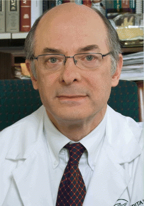
What needs to be in the follow-up of certain patients who have undergone treatment for thyroid cancer? Uncertainties still exist, but change is in the air. The 2009 American Thyroid Association (ATA) guidelines promise to clarify at least some issues that affect practice. Details were presented in a keynote talk given at the recent World Congress on Thyroid Cancer in Toronto by David Cooper, MD, Professor of Medicine and Endocrinology at Johns Hopkins University School of Medicine in Baltimore, who chaired the ATA 2009 Thyroid Cancer Guidelines Task Force.
Explore This Issue
October 2009Most patients with thyroid cancer present with isolated disease, or with lymph node involvement, but rarely do people present with more advanced disease, Dr. Cooper said. The recurrence rate ranges from 10% to 30%, depending on the nature of the tumor. People in the field assume that if recurrences are detected and treated sooner rather than later, morbidity and mortality would be decreased. However, evidence suggests that select low-risk patients may benefit equally from a watch-and-wait approach.
Use of Thyroglobulin Measurements
Detection techniques today, such as stimulated thyroglobulin (Tg) and high-resolution ultrasound, are far more sensitive than they were even two decades ago, meaning that recurrences (which many researchers consider as part of persistent disease) can be detected fairly early.
What is not clear at this point is whether the detection of these small-volume recurrences is really going to benefit the person if we find them sooner rather than later, he said. Many recurrences, if detected later, can still be treated effectively. Admittedly, some would not be as treatable, which is part of the dilemma pertaining to the value of early detection. A way to deal with this is to risk-stratify the patients, something recommended in the guidelines, he said.
A pivotal study published in 2001 showed that risk of recurrence corresponds strongly with age. When you’re young, the recurrence rates are high; when you’re old, they are high; and in the middle age range, your recurrence rates aren’t that high, he said.
 I think our guidelines have helped push along the concept that once a patient has been proven to be free of disease, it is no longer necessary to keep their serum TSH suppressed.
–David S. Cooper, MD
Along with age, the ATA guidelines add that the low-risk patient is one who has no detectable residual disease, has typical tumor histology, and no 131-iodine uptake outside the thyroid bed. Intermediate-risk patients have microscopic invasion of tumor in the perithyroidal soft tissues, whereas high-risk patients have macroscopic tumor invasion along with other evident risk factors.
Data are now emerging that confirm that many of the same risk factors that predict mortality also predict recurrence, Dr. Cooper said. Tumor size of more than 3 centimeters predicts lymph node recurrence rate, and the number of lateral nodes predicts recurrence rates in lateral nodes. Furthermore, the degree of extrathyroidal extension of the tumor, along with aggressive tumor histology, all indicate how aggressive the tumor is and can predict not only mortality but also recurrence.
European Standard Should be Used for Serum Tg Tests
But what about the use of serum Tg? There are a number of issues related to the use of serum Tg; to address some of these, the new ATA guidelines recommend using the European Tg standard. However, even when assays are standardized against this standard, there is still a lot of variability in thyroglobulin measurements among the various kits that are available at different institutions, he said.
Another hurdle is that anti-Tg antibodies occur in a larger percentage of thyroid cancer patients than in the general population. False negatives in immunometric assays and false positives in radioimmuno assays also continue to exist as problems. These are all areas that require further research to solve them, Dr. Cooper said. There can also be undetectable Tg, including stimulated Tg, in low-volume disease, in poorly differentiated tumors, and in people with previous radioiodine therapy for metastatic disease of the lymph nodes.
False positive Tg is important to note, and occurs in about 2% of patients. If a patient has a Tg of 10, or 20, or 30, and it cannot be found on imaging, the patient may end up being treated unnecessarily with radioactive iodine (RAI).
Another relevant issue in follow-up is that a rising Tg indicates that there is probably tumor progression, which may become clinically apparent at some point in the future, Dr. Cooper said. The new guidelines recommend that Tg be measured every six to 12 months by immunometric assay standardized against the European standard CRM 457, and it should be measured in the same laboratory using the same assay.
Additionally, periodic Tg measurement should be considered during follow-up with patients who have undergone less than a total thyroidectomy, and in patients who have not received RAI. This recommendation is new. We still think thyroglobulin can be useful in such patients, but we really don’t know what the cutoff should be in someone who hasn’t had a total thyroidectomy or RAI, Dr. Cooper said.
Use of Recombinant Human TSH
Dr. Cooper also discussed issues around stimulated Tg. A couple of recent pivotal papers showed that if Tg levels are low after stimulation, the chances of having residual disease or recurrence was also very low. But if Tg is high, there is an increased risk of recurrent disease. Recombinant human thyroid-stimulating hormone (rhTSH) testing has a similar pattern. Yet even if Tg levels are high after testing, there are still some people who do not have detectable disease using sensitive imaging.
In the ATA guidelines, low-risk patients who have had remnant ablation, negative cervical ultrasound, and undetectable Tg on suppression within the first year should get a Tg after thyroxine withdrawal or rhTSH in about a year after the ablation to verify the absence of disease, said Dr. Cooper. If the stimulated Tg is undetectable, patients are considered to be free of disease and need less intensive future follow-up.
Use of Various Imaging Modalities
In patients free of disease, the guidelines state that if the ultrasound and the stimulated Tg do not reveal any disease, we think you can follow the patient with a yearly clinical exam and Tg on thyroid hormone replacement rather than repeating the stimulated Tg, Dr. Cooper said.
He noted that the guidelines do not address the newly developed very-high-sensitivity Tg assays, which have a functional sensitivity of 0.05 to 0.1. These are new in the field, there is insufficient experience with them, and there are not yet any prospective studies using them.
Management of Tg-Positive, Scan-Negative Patients
With more sensitive tests, physicians now must deal with patients who have biochemical evidence of disease, with a detectable basal and/or stimulated Tg, but no evidence of any disease anatomically, despite our best efforts to find it, said Dr. Cooper. It is not known how best to manage these patients, although most seem to do well.
In general, a certain fraction of patients will have no clinical disease but will have a stimulated thyroglobulin greater than 1 or 2 ng/mL after recombinant TSH. Imaging will reveal disease in about a quarter or a third of such patients. The other two-thirds or three-quarters may have stable or decreasing thyroglobulin over time; it’s not necessarily a progressive thing, he said.
As for when it is reasonable to do imaging in patients, the guidelines suggest that it should be done with patients with a rising Tg of 10 ng/mL or more after thyroxine withdrawal or less than 5 ng/mL after rhTSH. The guidelines recommend ultrasound, high-resolution CT with contrast, or MRI. If imaging is negative, PET-CT should be considered. If PET is negative, physicians should consider empiric RAI therapy.
Generally, the role of PET scanning has expanded in the new guidelines, Dr. Cooper said. It is recognized as something that helps localize disease in Tg-positive, RAI scan-negative patients without needing to have a negative post-RAI therapy scan. PET scans should also be used for staging of disease, as a prognostic tool for patients with metastatic disease to identify those at highest risk, and for evaluation of post-treatment response.
The guidelines now emphasize risk-adapted management. Not only is the initial therapy directed toward the baseline risk of the patient, but the follow-up should also be based on the risk of the patient, Dr. Cooper said. This includes observing and acting on how risk changes over time in individual patients.
In low-risk patients, in the first two years of follow-up, it is now considered reasonable to obtain Tg levels every six months for the first two years. Once the stimulated Tg has been negative for a year, the test does not need to be repeated.
In patients with intermediate risk, it is reasonable to repeat stimulated Tg levels because disease will be detected in some of them-even if basal Tg is undetectable.
Ultrasound should be used yearly in low-risk patients for the first two years, but more frequently in intermediate- and high-risk patients (high-risk patients need to be intensively monitored in general). The guidelines now say that once a patient is disease-free, the serum TSH can be allowed to rise into the normal range in low-risk patients, but should remain <0.1 mU/L in intermediate- and high-risk patients. Dr. Cooper noted that the approach to thyroid hormone suppressive therapy needs to take into account other comorbidities a patient has (such as cardiovascular disease or osteoporosis) and factor those into the risk-benefit treatment profile for that patient.
I think our guidelines have helped push along the concept that once a patient has been proven to be free of disease, it is no longer necessary to keep their serum TSH suppressed, he said.
©2009 The Triological Society
Leave a Reply