Optical coherence tomography (OCT) provides a method to view tissues at the micron level, and for the delicate tissues of the vocal folds, this procedure offers much of the cellular information of a biopsy without the associated morbidity.
Explore This Issue
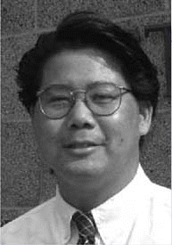
September 2006OCT is an emerging technology, which in many disciplines is still investigational, said Brian J. F. Wong, MD, PhD, Associate Professor of Otolaryngology-Head and Neck Surgery at the University of California-Irvine.
I think the most important thing that OCT of the head and neck allows you to determine is the integrity of the basement membrane, said Dr. Wong, who is also associated with the Department of Biomedical Engineering and The Beckman Laser Institute at UC-Irvine, and in the case of early cancers, when the integrity of the basement membrane is compromised, one’s index of suspicion with respect to early invasive cancer is extremely high.
There has been a gradual progression in the capabilities of this technology over time, Dr. Wong said. OCT is different from other imaging modalities in that it provides extremely high-resolution images which approach that of light microscopy.
OCT is an infrared imaging technology that is very similar in application to ultrasound, but instead of using sound waves, light waves are used.
Early Research, Promising Results
Dr. Wong and colleagues at UC-Irvine have been engaged in studies which have produced what is believed to be the largest clinical series of head and neck patients examined by OCT (Laryngoscope 2005;115:1904-1911).

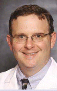
Detection of the disruption of tissues of the larynx basement membrane, which is the hallmark for invasive cancer, is one of the chief benefits of OCT, agreed William B. Armstrong, MD, Associate Professor of Clinical Otolaryngology at UC-Irvine. A colleague of Dr. Wong’s, Dr. Armstrong is also working on a prototype OCT device being developed by scientists and researchers at the Beckman Laser Institute.
Dr. Armstrong presented findings recently on his work with OCT and head and neck cancer patients at the Middle/Western Section of the Triological Society Meeting earlier this year in San Diego.
How OCT Works
OCT is an imaging modality that uses light to produce cross-sectional images of tissue, Dr. Wong said. This technology was developed by James G. Fujimoto, MD, and colleagues at the Massachusetts Institute of Technology (Science 1991;254:1178-1181).
OCT has a depth of penetration of up to three millimeters and the resolution is much finer than with other imaging modalities, Dr. Armstrong said. The diameter of a red blood cell is about eight microns, and the best you can get with CT scans would be a millimeter, which would be a hundred times less resolution, he added. CT, ultrasound, and MRI all have resolutions that are at least an order of magnitude less than that available with OCT. Current research OCT systems have resolutions as low 10 microns-and there are systems in development that will have resolutions as low as one to two microns.
I think the most important thing that OCT of the head and neck allows you to determine is the integrity of the basement membrane. – -Brian J. F. Wong, MD, PhD
OCT is an infrared imaging technology that is very similar in application to ultrasound, Dr. Armstrong said, but instead of using sound waves, light waves are used. The OCT procedure shines light into the tissue using infrared light at low power levels and then examines the backscattering that comes from the reflections to create a picture of the tissue structure.
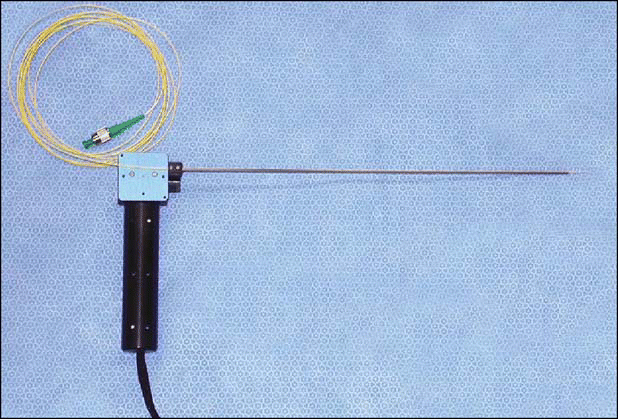
The OCT device uses a super-luminescent diode, Dr. Armstrong added, which produces infrared light. The device is passed down a small, approximately one-millimeter optical fiber that is housed inside a plastic sheath, which is in turn housed inside a metal tube.
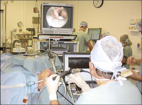
To deliver the OCT device, Dr. Wong said, an endoscope, a probe, a microscope, or a hand-held device can be inserted into the mouth. It really depends on the clinical application, but the best quality images are obtained using a probe system, a pencil-like device that can be placed in direct contact or near contact with the tissue, he added.
In most present experiments, Dr. Armstrong said, the device has been delivered through an endoscope to patients who are under general anesthesia. The ultimate longer term goal is to develop a way to get images and get data on a person who is awake in the office. This would involve having the patient hold still with the tongue protruded and would probably require a topical anesthetic.
What the Technology Provides
With the technology we have, we can see the architecture of the tissues, Dr. Armstrong said. OCT provides such details as the thickness of the epithelial layer, the location of glands and blood vessels, and disruption of the basement membrane.
OCT has incredible value for looking at thin-layered structures, Dr. Wong said. It is of extreme value in organ and tissue sites where a biopsy of that organ could lead to the destruction or severe functional impairment of the organ. Anything that is done to physically perturb the vocal cords can change the intrinsic modes of vibration, altering ultimately the quality of the voice, so having a high resolution imaging technology to look at the vocal cords has extreme utility and value. OCT may provide the ability to guide the decision to perform a biopsy and provide information on the most beneficial placement of the biopsy.
We are trying to identify and characterize both normal and pathological structures and we are still in the early stages in terms of optimizing the equipment in terms of ergonomics, of being able to use it in a non-invasive manner, and in terms of miniaturization. – -William B. Armstrong, MD
State of Present Research
At the present time, Dr. Wong said, one cannot go out and buy an OCT machine for use in body sites apart from the retina and the skin. OCT for the tissues of the head and neck is still an investigational technology-but is an extremely active area of interest in research in optical diagnostic technology.
There is no competition at this time for a non-invasive high-resolution imaging technique, he added, so in my opinion it is a matter of time, dictated largely by the demonstration of the value of this technology and obviously you would have to convince third-party payers that this is something that should be reimbursed. Convincing third-party payers can be very difficult, he added, as demonstrated by the relatively slow adoption of PET scanning, for example, which took 20 years to get reimbursed by Medicare. But we feel that this technology can make the same impact in medicine for superficial disease in thin structures as PET scanning has for localization of functional differences in tissue metabolism.
The research being done at UC-Irvine includes examination of the pediatric airway, looking at subglottic stenosis, designing devices for use with flexible endoscopy, and designing devices for use with surgical microscopes. The focus has been on instrumentation and nanotechnology applications, Dr. Wong said.
We’ve been able to see the morphology of Reinke’s edema, Dr. Wong added. We are seeing discrete fluid pockets in Reinke’s edema as described by pathologists a hundred years ago and it is fascinating to see these in vivo.
Dr. Wong’s clinical research protocol is not using OCT to alter patient care at this time-the goal is to determine the efficacy of this technology.
We are still at the stage where we are trying to learn the capabilities of the equipment, Dr. Armstrong said.
Limitations
There are limitations for the OCT procedure, Dr. Wong said. The tissue viewed must be a thin structure. There is a trade-off in all imaging technologies where one has to sacrifice resolution for depth of penetration or imaging-so the higher the resolution in general, the less deep you can visualize the tissue-on the other hand, getting information with respect to depth usually results in a loss of resolution.
OCT is not as good as microscopy, but you see much deeper in the tissue, on the order of about a millimeter, with very high resolution. Current systems in our laboratory are approaching one micron resolution, Dr. Wong added.
Future
This technology will likely follow the same adoption and usage dynamics as CT, ultrasound, and MRI, Dr. Wong said.

The OCT procedure will require training, Dr. Wong added. I think that this is the type of technology that is probably going to make its way into residency training programs first, but I think that clinicians who want to use OCT in their offices are probably going to take some CME courses.
With new imaging modalities, the line between surgeon, pathologist, and radiologist is blurring, Dr. Wong noted. OCT technology is certainly going to involve pathologists, but a pathologist is not in the operating room with the patient-there are other technologies that are being developed where surgeons are really going to have to understand how to do imaging. This is already happening in other applications-surgeons know how to interpret ultrasound so they can use the information intraoperatively or in the office or clinic. So there is certainly a precedent for that, Dr. Wong added.
More work still needs to be done in terms of instrumentation design and technology improvement, Dr. Wong noted. The capabilities of OCT need to be improved in terms of acquisition time speed and resolution. This is an active area of research by investigators in several institutions in optical sciences in the US, Europe, and Asia.
We are trying to identify and characterize both normal and pathological structures and we are still in the early stages in terms of optimizing the equipment in terms of ergonomics, of being able to use it in a non-invasive manner, and in terms of miniaturization, Dr. Armstrong said. Research work is also focusing on improving the software and hardware so that it can be easily used in a clinical situation and be adaptable to the clinician.
Research systems at UC-Irvine could be taken into clinical implementation in the next two years, the researchers predict.
©2006 The Triological Society


Leave a Reply