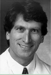
WASHINGTON, DC-Practice guidelines have recently been developed for patients with thyroid nodules and differentiated thyroid cancer. David S. Cooper, MD, Director of the Division of Endocrinology at Sinai Hospital and Professor of Medicine in the Division of Endocrinology at Johns Hopkins University School of Medicine in Baltimore, who chaired the committee that developed the guidelines, participated in a panel discussion about the guidelines at the recent AAO-HNS Annual Meeting.
Explore This Issue
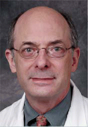
December 2007 David L. Steward, MD, said that ultrasound-guided fine needle aspiration can be performed in the office at a patient’s initial visit.
Scope of the Problem
Thyroid nodules are common-present in about 5% in women and 1% in men living in iodine-sufficient parts of the world. However, the prevalence is likely higher because high-resolution ultrasound can detect thyroid nodules in 19% to 67% of randomly selected individuals. The clinical problem is to isolate the 5% to 10% of these nodules that are malignant.
Differentiated thyroid cancer, which includes papillary and follicular carcinoma, comprises the vast majority (90%) of all thyroid cancers in the United States-about 33,500 cases predicted in 2007, a number that is slowly and steadily increasing.
Over the past decade there have been many advances in diagnosis and treatment of thyroid nodules and differentiated thyroid cancer. Controversies exist about the most cost-effective approach to diagnosis, the extent of surgery for small cancers, use of radioactive iodine to ablate remnant tissue after thyroidectomy, appropriate use of thyroxine suppression therapy, and the role of recombinant human thyrotropin.
For all these reasons, the American Thyroid Association formed a task force to examine current strategies and to develop clinical guidelines using principles of evidence-based medicine. (Management guidelines for patients with thyroid nodules and differentiated thyroid cancer. Thyroid 2006 Feb;16(2):109-42, available online at www.thyroid.org .)
Thyroid Nodules
A thyroid nodule is a discrete lesion within the gland that is palpably and/or ultrasonographically distinct from the surrounding thyroid parenchyma. (However, nonpalpable nodules have the same risk of malignancy as palpable ones of the same size.) Generally, only nodules larger than 1 cm should be evaluated, because they have the potential for clinically significant cancers. Appropriate evaluation consists of:
- History and physical exam, focusing on the thyroid gland and adjacent cervical lymph nodes, and looking for a history of head and neck irradiation, total body irradiation, first-degree family history of thyroid cancer, and exposure to nuclear fallout.
- Serum thyrotropin (TSH). There is some evidence that high preoperative TSH may predict a higher sensitivity for postoperative surveillance with TSH, but it is not known how this would affect patient management.
- If TSH is suppressed, radionuclide thyroid scan to determine if the nodule is autonomously functioning, and therefore most likely benign.
- Thyroid ultrasound to determine if the size and location of the nodule corresponds to what was discovered on palpation, as well as to see if there are other nonpalpable nodules.
- Serum calcitonin measurement to detect early C-cell hyperplasia and medullary thyroid cancer is not recommended in the current ATA guidelines because of lack of data on cost-effectiveness.
- FNA biopsy, the most accurate and cost-effective method to evaluate thyroid nodules. Results are divided into four categories: benign (the most common reading), indeterminate or suspicious for neoplasm, nondiagnostic, and malignant (5-10%).
Patients with multiple thyroid nodules have the same risk of malignancy as those with solitary nodules, and a diagnostic ultrasound should be performed to delineate the nodules. Nodules with suspicious sonographic features (hypoechoic, calcifications, indistinct borders, high blood flow on Doppler) should be biopsied first. A new study presented at the AAO-HNS meeting showed that in 447 office-based fine-needle aspiration (FNA) biopsies of the thyroid gland (using ultrasound-guided fine needle aspiration, or US-FNA), more than 92% resulted in an adequate specimen, thus saving patients a visit to the hospital for the procedure. These findings suggest that US-FNA can be performed in the office at a patient’s initial visit, said the study’s lead author, David L. Steward, MD, Associate Professor in the Department of Otolaryngology and Director of the Clinical Trials Program at the University of Cincinnati.
Patients with benign thyroid nodules require follow-up because of a 5% rate of false negative FNA results. Although benign nodules may decrease in size, they do so slowly, and size is not an indication of the potential for malignancy. Follow-up should consist of serial ultrasonography.
In terms of medical therapy, routine suppression of serum TSH for benign thyroid nodules is not recommended, but such nodules can be excised if they enlarge and if there is reason for clinical concern.
For indeterminate biopsies, surgery is generally recommended, since the rate of malignancy is 15% to 20%. Lobectomy is appropriate for patients who prefer a limited procedure. For a large tumor, total thyroidectomy is indicated to prevent the need for reoperation in the event the tumor is malignant.
Nondiagnostic biopsies need to be repeated, preferably with ultrasound guidance. Cystic nodules that repeatedly yield nondiagnostic aspirates need close observation or surgical excision. For repeatedly nondiagnostic biopsies of solid nodules, surgery or observation are both appropriate.
For a biopsy diagnostic of malignancy, near-total or total thyroidecomy is the procedure of choice, unless (1) the tumor is less than 1 cm in diameter and completely confined to the thyroid, (2) there is no evidence of ipsilateral adenopathy on ultrasound, and (3) there is no family history of thyroid disease or history of head and neck irradiation. In such cases, lobectomy may be appropriate. Some experts, including the American Thyroid Association, recommend routine central neck dissection for patients with biopsy-proven thyroid cancer, but this remains controversial.
 David S. Cooper, MD, chaired the committee that developed the guidelines for patients with thyroid nodules and differentiated thyroid cancer.
Differentiated Thyroid Cancer
Goals of differentiated thyroid cancer therapy include:
- Removal of the primary tumor, disease that has extended beyond the thyroid capsule, and involved cervical lymph nodes.
- Minimization of treatment- and disease-related morbidity.
- Accurate staging.
- Postoperative treatment with radioactive iodine, where appropriate.
- Accurate long-term surveillance for disease recurrence with radioiodine whole-body scanning and measurement of serum thyroglobulin.
- Minimization of the risk of disease recurrence and metastatic spread, most importantly by means of adequate surgery and adjunctive treatment.
Preoperative staging should be done by neck ultrasound, because 20% to 50% of papillary carcinomas spread to cervical lymph nodes. Frequency of micrometastases can be as high as 90%, and preoperative ultrasound can identify suspicious cervical adenopathy in about 25% of cases. Moreover, accurate staging is important in determining prognosis and tailoring individual treatment. However, the presence of metastatic disease does not obviate the need for surgical excision of the primary tumor, because metastatic disease can respond to radioiodine therapy. In such cases, because locoregional disease is an important component, it is important to remove the entire thyroid gland in addition to the primary tumor. The American Thyroid Association guidelines do not recommend routine neck CT or MRI for preoperative evaluation. Iodinated contrast should be avoided, as it will interfere with postoperative radioiodine therapy.
Postoperative staging for thyroid cancer-as for all cancers-is used to:
- Establish a prognosis.
- Tailor decisions about postoperative adjunctive therapy, including radioiodine and thyrotropin suppression.
- Make decisions about the frequency and intensity of follow-up.
- Enable accurate communication between patients and health care professionals.
Application of the AJCC/UICC classification system based on pTNM parameters is recommended for tumors of all types because it provides a useful shorthand method to describe the extent of the tumor. It is also used for hospital cancer registries and epidemiologic studies. It is recommended for all patients with differentiated thyroid cancer. Postoperative clinicopathologic staging systems also are recommended to improve prognostication and to plan follow-up.
©2007 The Triological Society
Leave a Reply