Endoscopic surgery provides a less invasive and highly visual approach to skull base tumors and can reduce morbidity compared with open surgery, according to the experts interviewed for this article. While the role of endoscopy continues to evolve as surgeons treat increasingly larger and more difficult skull base lesions, a number of contraindications and precautions need to be kept in mind.
Explore This Issue
November 2007An Evolution
Endoscopic skull base surgery has evolved from endoscopic sinus surgery, explained Brendan Stack, MD, Vice Chairman of the Department of Otolaryngology–Head and Neck Surgery, and Director of the Divisions of Head and Neck Oncology and Clinical Research at the University of Arkansas for Medical Sciences.
“It’s gone from taking care of sinus disease, to repairing brain fluid leaks, to removing small benign lesions in the skull base, to removing increasingly larger benign lesions, as well as many malignant lesions,” he said.
Endoscopic skull base surgery developed from using this technique to treat sinus inflammatory disease, orbital decompressions, epistaxis, and cerebrospinal fluid (CSF) leaks and to conduct endoscopic biopsy, said Martin J. Citardi, MD, a Cleveland Clinic Head and Neck Institute rhinologist.
Now, skull base indications are evolving across time and are different at various institutions, said Dr. Citardi. Overall, the medical community is realizing that endoscopic approaches are a viable treatment strategy for the management of skull base neoplasms, he said.
Skull Base Indications Growing
Endoscopic skull base surgery is most effective for smaller growths, although surgeons are pushing these limits and approaching lesions of different sizes, said Dr. Stack.
For example, juvenile nasal angiofibromas are large tumors filled with blood vessels, which, prior to improved endoscopic optics and ways of achieving hemostasis, needed open surgery for removal, said Dr. Stack. “Five years ago or more, those patients were destined to have an open operation because they would bleed so much,” he said.
Size is not necessarily a limiting factor for removing tumors, said Pete Batra, MD, Assistant Professor of Surgery at the Cleveland Clinic Head and Neck Institute.

“The earlier literature has focused on smaller tumors because as you accrue experience you start with smaller growths and tackle increasingly larger tumors as time goes on,” he said. “We can now remove much larger tumors that are accessible by endoscope.”
In addition to tumor size, its type or histology often do not affect how the disease is approached endoscopically, said Dr. Citardi. Regardless of tumor type, its removal is usually the same, he said.
Carl Snyderman, MD, professor of otolaryngology and neurological surgery and co-director of the Center for Cranial Base Surgery at the University of Pittsburgh School of Medicine, uses an endonasal approach for tumors involving the ventral skull base below the anterior brain or arising in the nasal passages. Examples include olfactory neuroblastomas, meningiomas, pituitary tumors, angiofibromas with intracranial extension, and inverted papilloma with skull base involvement.
Treating malignant nasal tumors that involve the skull base and intracranial tumors, both benign and malignant, can be controversial, added Dr. Snyderman. Some physicians express concern about using the procedure for malignancies because oncologic principles need to be applied to these growths to achieve complete resection without compromising margins.
However, a multidisciplinary team approach can ensure appropriate management of these tumor types, he explained, adding that only a handful of institutions are performing these surgeries.
Advantages
One advantage of endoscopic skull base surgery is that it is minimally invasive, said Dr. Batra. Additionally, the rigid endoscope is a tool that provides brilliant illumination and magnification to detect tumor origin and boundaries and allows surgeons to see around corners. The endoscope allows superb exposure to the sphenoid sinuses, frontal recess and frontal sinus, and the orbital apex, said Dr. Batra, adding that this, in turn, aids with precise tumor removal.
The endoscopic approach to skull base surgery improves visualization, agreed Dr. Snyderman. However, he views the procedure as being maximally, not minimally, invasive. “The endoscope has extended the limits of skull base surgery, and we’re getting to areas we couldn’t get to before,” he said. “It’s maximally invasive but avoids the morbidity of extracranial approach.”
When compared with open skull base procedures, the endoscopic approach not only reduces morbidity but also provides similar overall survival and disease-free survival in published reports, said Dr. Citardi. Furthermore, endoscopic techniques are associated with less time in the hospital for the patient, he added.
In many cases, the endoscopic approach can also provide cosmetic benefits versus open surgery, said Dr. Snyderman.
Moreover, larger skull base surgeries being performed endoscopically help to reduce the need for some free flaps, said Dr. Stack. These procedures leave more local tissue available for reconstruction, and these defects have also been successfully repaired with allografts, he said.
Even though the endoscopic approach to the skull base may have advantages, physicians need to use the surgical approach that is most appropriate for the patient’s disease, said Dr. Snyderman. “Going through the nose is sometimes a better approach than going through the side of head and through the cranium,” he said. “It should be individualized for the patient.”
Contraindications, Precautions, and Limitations
Although endoscopic skull base surgery is advantageous for a variety of indications, the procedure also has a number of contraindications and should be approached with several precautions in mind, agreed the physicians interviewed for this article. The surgery also has some limitations and a few disadvantages when compared with open procedures.
Contraindications
Extremely vascular tumors are a relative contraindication, and whether or not to perform the procedure depends on the technical expertise of the physician, said Dr. Batra.
The procedure is also contraindicated for extensive tumors that involve the facial soft tissues, Dr. Batra said. A combined open and endoscopic approach may be possible, but this sort of tumor is most often amenable to an open procedure, he explained.
If the tumor is a massive bilateral growth, removing it with endoscopic skull base surgery also depends on the surgeon’s technical expertise, said Dr. Batra.
Additionally, if a skull base tumor involves orbital contents, endoscopic techniques alone are inadequate in most instances, since orbital exenteration will be required, said Dr. Citardi. However, using a combination of open incision and visualization with an endoscope may be possible, he said.
Overall, if the tumor is inoperable by traditional methods, it is not a candidate for curative resection by endoscopic skull base surgery, said Dr. Batra.
Precautions
Surgeons performing endoscopic skull base surgery should be concerned about approaching critical areas such as the orbit and optic nerve, brain and surrounding dura, and the internal carotid artery, said Dr. Batra.
Endoscopic surgeons also need to be aware of complications such as bleeding, which can be life-threatening, as well as the potential for blindness, brain injury, and spinal fluid leakage, said Dr. Citardi. “That’s why preoperative assessment is very important,” he said. “Patients need to undergo specific imaging and pretreatment planning with a multidisciplinary team.”
To help plan surgery and avoid sensitive structures, physicians view computed tomography (CT) and magnetic resonance images of bony and soft tissue before surgery to map out the sinuses from the eyes to the skull base in three dimensions, said Dr. Batra. “This navigation also helps physicians determine if they are near any critical blood vessels or other structures,” he said.
Intraoperative CT scanning is a new modality that may allow physicians to assess resected areas and determine if any residual tumor remains, said Dr. Batra. “At an early juncture, we have found this modality to be a helpful adjunct for tumor resection.”
Limitations and Disadvantages
When compared with open skull base surgery, an endoscopic procedure may take longer, noted Dr. Snyderman. “It can be physically and emotionally exhausting to perform the procedure for hours on end,” he said.
In addition, there may be a higher incidence of small postoperative CSF leaks because reconstruction using endoscopy is more difficult than reconstruction with an open procedure, said Dr. Snyderman. “You have a better opportunity for watertight reconstruction with open surgery,” he said.
In the nose, surgeons have to use a multilayer technique that is not watertight, he explained. Consequently, physicians will often use packing to exert pressure on the reconstruction to encourage tissue healing. A nasal septum mucosal flap, however, can cover openings leading to the cerebrospinal region and can improve healing, said Dr. Snyderman.
The level of technical expertise required to perform skull base endoscopic procedures can also be a limitation, said Dr. Batra. “You also have to have the necessary equipment, usually available at a tertiary care center, and a multidisciplinary team,” he said.
Another limitation is this procedure has a shorter track record compared with traditional operations, said Dr. Batra. “All centers are trying to accrue greater experience to make sure endoscopic skull base surgeries are as good as open approaches,” he said.
There are still a lot of missing data on outcomes for various tumor types using endoscopic skull base surgery, agreed Dr. Snyderman, adding that it will be a number of years before data show whether open and endoscopic skull base surgeries are comparable or if one is better than the other. “Early results suggest that the endoscopic procedure is a just as good with less morbidity,” he said.
Team Approach Needed
To help ensure the best outcomes of endoscopic skull base surgery, some institutions have developed a team approach to the procedure, said Dr. Stack. These teams will generally include neurosurgeons and otolaryngologists, as well as rhinologists, medical oncologists, and radiation oncologists, according to the experts interviewed for this article.
Despite the need for a multidisciplinary approach, individual otolaryngologists–head and neck surgeons perform the majority of procedures, said Dr. Stack. Rhinologists also perform sinus surgery and have developed the ability to do more complex procedures over time, he added.
One reason why a neurosurgery/otolaryngology team approach may not be available is that neurosurgeons often don’t use endoscopes, and there is some resistance within this field to become part of an endoscopic skull base surgery team, noted Dr. Stack.
“There is still some resistance in neurosurgery circles to a team approach,” agreed Dr. Snyderman.
Originally, many traditional head and neck cancer surgeons also did not like the idea of endoscopy, noted Dr. Citardi. “But in the last two to four years, there’s been a tipping point,” he said.
Whatever the medical background of the team, all experts interviewed for this article agreed that extensive training and an understanding of cranial base anatomy is necessary before physicians perform endoscopic skull base surgery.
“One of my concerns is that individuals who have tremendous experience with traditional techniques will go crashing ahead with endoscopic resection and may discredit the approach,” said Dr. Citardi.
The Future
The outlook for endoscopic surgery involves tackling increasingly difficult tumors and potentially adding robotics to these procedures, said Dr. Stack.
“There’s always an evolution of surgical techniques and technology to better take care of patients,” said Dr. Batra. “It’s conceivable that in the future robotic surgery may play a role, but it hasn’t been defined to date,” he said, adding that the technology is still in its early stages.
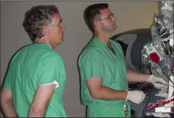
In 2005, Bert W. O’Malley Jr., MD, and Gregory Weinstein, MD, in the Department of Otorhinolaryngology–Head and Neck Surgery at the Hospital of the University of Pennsylvania, started the world’s first robotic skull base surgery program.
“So far, we have developed novel skull base approaches and surgical procedures in preclinical investigations, and then we have applied these robotic techniques to three patients with skull base benign neoplasms,” said Dr. O’Malley, Gabriel Tucker Professor, Department Chair, and Co-Director of the Head and Neck Cancer Center and the Center for Cranial Based Surgery.
Robotic-assisted head and neck and skull base surgery combines the technical and optical advantages of robotics with the minimally invasive advantages of classic endoscopic sinus and skull base surgery, said Dr. O’Malley. The robot has the potential to overcome many of the limitations and disadvantages of classic endoscopic skull base surgery such as the lack of bimanual dexterity, he added.
For example, in present endoscopic skull base surgery, surgeons must hold a scope or use cumbersome mechanical arm scope holders, while working through small openings in the nose or oral cavity. At the same time, the surgeon is attempting to manage secretions, control bleeding, resect the actual disease or tumors, and then repair skull base defects, he said.
“With open skull base surgery, you are able to use two hands or more with assistants in the operating room,” he said. “You also have top-down visualization and relatively prompt control of any bleeding. This is not readily possible with present endoscopic skull base surgery approaches and techniques.”
Robotic-assisted skull base surgery, on the other hand, provides manual dexterity equal to or better than open surgery, while working through small openings in the oral cavity and soon through the nose as the robotic instruments continue to be miniaturized, explained Dr. O’Malley.
Moreover, the robot has four arms that one person operates, he explained. “It provides amazing three-dimensional visualization, bimanual and even trimanual dexterity, fine instrument mobility, and overall precise dissection,” he said.
While beneficial, the robotic itself and associated instruments are expensive, and as with any technology, there is the possibility of mechanical failures, said Dr. O’Malley. There is also a learning curve involved with using the robot; however, Dr. O’Malley and Dr. Weinstein believe that this curve is much easier than that needed to learn other transoral and endoscopic skull base surgical techniques.
The most apparent limitation of the robot in skull base surgery or even standard endoscopic sinus surgery is that current instrumentation is not designed for endoscopic or transoral minimally invasive skull base surgery, said Dr. O’Malley. “We’re working hard to adapt it for skull base surgery,” he explained.
Dr. O’Malley and Dr. Weinstein, in addition to the early adopter surgeons whom they have trained over the past year, are hoping to introduce much smaller instrumentation in the near future, which should enable more widespread use in skull base surgery.
How soon such technology is available depends on funding from medical device companies, said Dr. O’Malley. “It could take six months to six years,” he said. “Overall, I strongly feel that this is the wave of the future for head and neck and sinus and skull base surgical procedures,” he concluded.
©2007 The Triological Society


Leave a Reply