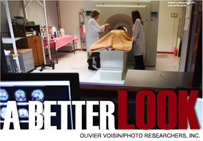
When it comes to treating head and neck tumors, the more information that is available, the better. In the past, options for investigating these types of tumors and their aggressiveness were limited. But advances in optical imaging, positron emission tomography (PET) scanning, magnetic resonance imaging (MRI) and fluorescent and ultrasound imaging have some otolaryngologists excited about the prospect of getting a better look at head and neck cancer.
Explore This Issue
April 2011“I think all of us surgeons are frustrated when we see patients and we treat them with what we think is a very good treatment paradigm only to see them not do as well as we think they should do for reasons that we really don’t understand,” said Mark Wax, MD, professor of otolaryngology-head and neck surgery at Oregon Health and Science University in Portland. “I think all of us are excited by all of the time and all of the research that’s going on that may … allow us to stratify our patients into different treatment paradigms that will decrease their morbidity and increase their chances for good functional outcomes.”
PET/MR
A scanner that combines the molecular imaging of PET and magnetic resonance (MR) was recently approved for commercial use in Europe and is expected to become available soon in the U.S., possibly later this year.
The scanners could become a must-have, used as commonly as PET/CT scans but with added advantages: MR is better at imaging soft tissue and doesn’t involve the radiation of a CT scan. The technology combines the ability of the PET scan to highlight biological processes with the superior structural display of MR technology.
“If you don’t have anatomical co-registration for that (PET scanning), you don’t know whether that’s a normal structure,” said Kurt Zinn, DVM, PhD, director of advanced medical imaging research at the University of Alabama at Birmingham. “You can’t really interpret it unless you have the anatomy together with it.”
PET/MR promises to outperform the PET/CT combo, he said. “Especially in the head and neck area, the MR is much better at imaging of nerves and the brain and those sorts of things, relative to CT,” he added. Dr. Zinn said he expected quick adoption of the technology in the U.S., possibly in 2011, despite the price tag of $4 million to $5 million.
A possible question for PET/MR scanning will be who performs the scans. Physicians certified in nuclear medicine typically perform PET scans but are not trained in MR technology, Dr. Zinn said. Radiologists are trained and certified in both PET and MR and can read both. “There will likely be some turf battles and adjustment until equilibrium is reached,” he said.
Dr. Zinn added that interpretation of MR images in PET/MR scanning will be more difficult than CT was for PET/CT scanning. “Some feel nuclear medicine physicians will be replaced by radiologists in the future,” he said in an e-mail. “Nuclear medicine was able to add CT training, but that will be harder to do for MR.”
Finding enough board-certified radiologists to read PET/MR scans may be one early challenge, he said.
New PET Tracers
PET scanning has been limited by the availability of the tracers used to detect tumors. The most commonly used tracer, fluorodeoxyglucose, or FDG, highlights areas of glycolysis, which is heightened in tumor cells. FDG can lead to false positives, however, because glycolysis is also heightened during inflammation and tissue repair.
New tracers are being developed to tailor to other metabolic activities that could give insights into what is happening in cancer cells, Dr. Wax said, citing a study from the Journal of Nuclear Medicine (2010;51:66-76).
“Every cancer is not the same, and with improved probes to image specific processes, we can tailor our therapies and treatments to the individual patient,” Dr. Zinn said. “Right now we can treat tumors differently depending on certain classification. But we can probably do a lot better at characterizing them by using molecular markers to help us know exactly how to treat, and, more importantly, during the treatment, to evaluate whether the tumor’s changing.”
Immuno-PET tracers, in which specially formulated antibodies are particularly reactive with a receptor that is more highly expressed in tumor cells, is an area of great promise, Dr. Wax said.
Epidermal growth factor-based and vascular endothelial growth factor-based antibodies are among those being used in these tracers.
18-F-FLT (18-F fluorodeoxythymidine) and 18-F-FMISO (18-F fluromisonidazole) are two of the more promising agents. FLT would help assess tumor proliferation, and FMISO is used to gauge hypoxia.
“It’s possible that we may be able to target specific areas of tumor sites, where they’re undergoing metabolic changes, parts of the tumor that are hypoxic, parts of the tumor that are showing angiogenesis, and then be able to target our therapies towards those areas,” Dr. Wax said. That’s really abstract, pie-in-the-sky type things, but I think that’s along the lines of what we’re trying to do.”
Dr. Wax emphasized that the new tracers are still only the subject of lab work, and the cost is an unknown. Any training would involve the interpretation and preparation of the materials, but how long that training would take is also undetermined, he said.
Fluorescence Imaging
Another area showing some promise uses fluorescence to decipher what is going on within tumors. In fluorescence imaging, a laser can be used to excite fluorophores, molecular components that absorb and emit light. Sensors can then capture the amount of fluorescence being emitted.
“We can correlate that amount of fluorescence with pathology that we obtained at biopsy,” said Gregory Farwell, MD, associate professor of head and neck oncology-skull base surgery at the University of California at Davis. “What’s become apparent in our studies (Arch Otolaryngol Head Neck Surg. 2010;136(2):126-33) and other groups’ studies is that there’s a very nice correlation between the fluorescence of a tissue and its pathologic state.”
The technology can help make diagnosis a simpler process, Dr. Farwell said. “It can allow for essentially a non-invasive biopsy,” he said. “In areas that are suspicious, we can analyze them with a probe and determine the composition of a tissue without a biopsy.”
In India, with the help of a grant from the Bill and Melinda Gates Foundation, the technology is being tested for the rapid assessment of large numbers of people.
“They’re looking at the ability of this technology to do mass screenings and improve health care delivery to patients at risk for head and neck cancer,” Dr. Farwell said.
Dr. Zinn said that potential application in the operating room is driving the development of fluorescence, particularly in robotic surgery, because the technology could easily be integrated into the tools already used.
“Fluorescence has a much higher spatial resolution than the MR or PET,” he said. “And, of course, it has the ability to zoom in to very small areas and detect sub-millimeter disease potentially. Even 50 to 100 tumor cells could be detected if you had the contrast to show that those cells are there. And you would never be able to do that on a PET- or MR-based approach.”
Dr. Zinn added that more probes are needed to maximize the potential of fluorescence imaging. That includes development of “quenched” probes, for which there is no fluorescence until the tracer is activated by a process in the cancer, offering better visibility by effectively lighting up only the disease.
There are clinical trials afoot for these advances in tracers for fluorescence imaging, but the work has been slow going, Dr. Zinn said.
Ultrasound Imaging
Ultrasound imaging is being refined with the use of tiny gas bubbles, contrast agents developed to enhance the acoustic signature of blood and to better define other structures in the body. In an assessment of thyroid nodules, for example, ultrasound with contrast bubbles determined benign or malignant status with sensitivity and specificity between 83 percent and 94 percent (Thyroid. 2010;20(1):51-57).
Because existing ultrasound machines can be used, the cost is relatively low at about $125 per exam, with just the extra cost of the microbubbles.
Kenneth Hoyt, PhD, assistant professor of radiology at the University of Alabama at Birmingham, said his group has been working to refine the process of coating microbubbles with antibodies that bind only to biomarkers in tumor vasculature.
“This technique can enhance signal specificity at tumor sites and promises such things as cancer detection at earlier stages where successful treatment is much more likely,” Dr. Hoyt said.
Microbubbles have also been found to help with drug delivery; the mechanical stresses of the procedure can temporarily increase the permeability of the cell membrane.
“It is essentially a lipid- or protein-shelled microbubble with a hollow gas core,” Dr. Hoyt said. “You could imagine loading that microbubble with some drug. Since these microbubbles are sensitive to ultrasound exposure, you can actually burst them at the tumor site to maximize drug delivery. Once again, this is a promising technology that we are on the verge of hearing much more about.”
The familiarity of ultrasound imaging might bode well for its future use with these refinements in place.
Randal Weber, MD, professor and director of surgical services in the department of head and neck surgery at the University of Texas’ M.D. Anderson Cancer Center in Houston, said surgeons are always looking for improved imaging that will delineate the extent of disease and facilitate surgical planning.
“For those of us that do tumor surgery, readily available 3-D imaging that’s not too resource- or labor-intensive could be useful for tumors of the skull base and parapharyngeal space,” he said. “It helps you plan an operation. Although radiologists can readily reconstruct 2-D images into 3-D images in their mind, it’s harder for those of us who don’t do that very often or don’t think that way.”
Narrow-Band Imaging
In this technique, light is restricted to just blue and green wavelengths, helping to highlight areas of increased vascularity that can indicate possible tumor tissue.
“One of our frustrations is that we often miss small tumors, or, in patients that have had radiation, it can be very difficult to evaluate because the tissue has been damaged by the radiation,” Dr. Farwell said. “And any tools that we have to make tumors more obvious will increase our sensitivity in picking up these tumors at an earlier stage.”
Studies have shown that the imaging works to improve contrast of vasculature (Eur Arch Otorhinolaryngol. 2010;267(3):409-14; Auris Nasus Larynx. 2009;36(6):712-716). Still, Dr. Farwell said more work is needed for the technology to prove itself. “The data on narrow-band imaging is pretty early,” he said. “It will be interesting in the next several years [to see] how people incorporate this into their practice.”
Machines with narrow-band imaging cost several thousand dollars.
Worth the Investment?
When it comes to new technology, the tough decision is always whether or not to invest.
“Cost is always a factor,” Dr. Wax said. “You don’t want to put all your eggs in one basket. But neither do you want to be investing too little in too many areas.”
Often, the choice comes down to seeing whether a technology will get reimbursed, Dr. Weber said.
“Ultimately, if you’re going to put an image modality into place that’s going to be used routinely on patients, there has to be a mechanism to get paid for it. So you’ve got to transition from it being a research tool to a clinical tool that’s valuable.”
Even in cases in which the investment is significant, it may be worth it, Dr. Farwell said. “If we can find tumors earlier and treat them at an earlier stage,” he said, “that’s not only cost effective but incredibly effective in helping our patients beat their disease.”
Leave a Reply