TORONTO-Image-guided sinus (IGS) and skull base surgery is no longer considered experimental or investigational, and is appropriate for use by otolaryngologic surgeons to help clarify complex anatomy encountered during functional endoscopic sinus and skull base surgery (FESS). However, although it is a state-of-the-art approach, it is not in of itself the standard of care.
This was one of the messages from otolaryngologists who spoke at the panel Image-Guided Sinus Surgery: State of the Art at the recent annual meeting of the AAO-HNS. Topics discussed by panel members included standard of care, a description of the evolution of the technology, types of cases where IGS helps, and glimpses of a virtual endoscopic sinus surgery simulator.
When Should IGS Be Used?
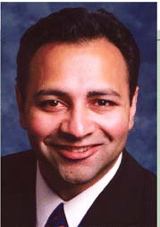
When it comes to IGS, there are some cautions. You should not feel that navigation is mandatory in all your cases, and you should feel comfortable that standard of care is what you provide patients, and what you feel is safe, said Sanjay Parikh, MD, Assistant Professor of Otorhinolaryngology-Head and Neck Surgery and Pediatrics at the Albert Einstein College of Medicine in New York, who moderated the panel and presented a talk relating to IGS and standard of care.
Although few otolaryngologists used IGS only five or six years ago, the technology is commonly found in practice now, Dr. Parikh said. It is used to identify complex anatomy in procedures such as revision sinus surgery, distorted sinus surgery, extensive sino-nasal polyposis, surgery relating to disease that abuts the skull base, optic nerve or carotid artery, and more.
The AAO-HNS has guidelines and a position statement for the use of IGS; these show that the technology is no longer considered experimental.
There is sufficient expert consensus opinion and a literature evidence base to support its use. However, it’s impossible to corroborate this with level one evidence, Dr. Parikh said. Studies in the literature tend to be observational and are of small series of patients, meaning the evidence is weaker than that from randomized controlled trials.
However, the AAO-HNS endorses the intraoperative use of computer-aided surgery in appropriate select cases to assist the surgeon in clarifying complex anatomy during sinus and skull base surgery, he said.
IGS provides a number of advantages in that it can provide added safety, makes FESS easier since landmarks are better defined, and is something that patients may prefer.
At the same time, it is not the standard of care, he said. And standard of care is making sure to provide a level of care and skill that is considered acceptable and appropriate by the medical profession.
Surgeons should be familiar enough with the anatomy in routine cases that they don’t need IGS, or can continue surgery if the IGS system goes down, Dr. Parikh said. IGS shouldn’t be used unless necessary.
Where the standard of care can fall down is if the surgeon doesn’t perform the appropriate diagnostic workup-such as not doing a sinus CT prior to sinus surgery-or if there is inappropriate medical treatment or a failure to discuss the risks, benefits, and alternative to surgery with the patient.
IGS can help you identify and confirm landmarks that you should be aware of. But you have to know what to do when you get there, Dr. Parikh said. Also, use of IGS needs to be well documented, clarifying medical necessity.
History of IGS
Richard Lebowitz, MD, Assistant Professor of Otolaryngology at the New York University School of Medicine, described some of the history behind the development of IGS. Original devices developed decades ago required rigid fixation of the head and were trajectory-based.
By the mid-1980s, frameless stereotactic surgery came into play, and had a position-sensitive articulated arm (wands). In the mid-1990s IGS progressed to having optical localizers, electromagnetic tracking, and were frameless, allowing for head mobility. At this point, the technology also no longer required use of wands, allowing for greater mobility of instruments.
Then there was a boom in technology, driven by surgeons’ needs, Dr. Lebowitz said. Recent years have seen several advances in endoscopic surgery devices. Now, IGS is used in revision surgery, extended frontal sinus surgery, tumor resection, skull base surgery, endoneurosurgery, and more.
Surgical instruments have become more refined as well. There are straight or curved microdebriders and drills, sinuplasty balloons, and frontal sinus and neurosurgical instruments.
Along with these are advances in CT, MRI, ultrasound, and fluoroscanning, each of which offers ever increasing details. Added to this, there is now the fusing of images, 3D volume rendering, and virtual image updates that are continually getting faster. Versions of these imaging devices are smaller and mobile, and can also be used intraoperatively.
IGS instrumentations now include compatible microdebriders-drills, universal instrument adapter systems, and even flexible instruments such as catheters and stents that can be used with hollow core sensors. In addition, ENTs have rotatable curved suction that does not require recalibration as the tip position changes.
We’ve seen big changes over the past decade, the next ten years will see even more more dramatic changes, said Dr. Lebowitz. One area to watch for is the development of robotic arms that can be used in conjunction with IGS. In future, it may open the door to things such as remote FESS, where the arm and patient are in one city, and the surgeon in another.
Limitations
The endoscope is a great instrument for us, but it’s not perfect, cautioned Brent Senior, MD, Chief of Rhinology, Allergy, and Sinus Surgery at the University of North Carolina, Chapel Hill. One difficulty lies in limits in the perspective the surgeon gets-along with problems of orientation and distortion.
We can often accept these distortions, but as we’re getting more into neuro-related surgery, distortion can be a problem, he said. For instance, more technology is needed for neurorhinology.
How Much Technology Is Necessary?
But at what point do otolaryngologists really need more technology? It’s tempting to become reliant on technology and imaging, but doctors still need their own clinical judgment.
Trust yourself, don’t trust a machine. Use IGS to confirm what you already know, not to figure out what you don’t know, Dr. Senior said. Indeed, it’s the skill of the endoscopist that determines whether or not a case even is performed endoscopically, not simply because one has IGS.
Dr. Senior provided examples of cases where IGS helped. One was of a 24-year-old man who underwent an endoscopic craniofacial resection for a large benign bony lesion involving the sinuses and skull base with significant intracranial extension. Here, IGS was useful because it allowed endoscopic drilling of the lesion in the setting of very distorted, even absent, landmarks.
A second case was a 62-year-old man who presented with altered mental status and was found to have a large pituitary neoplasm with spurasellar extension and ventricular compression. He underwent minimally invasive pituitary surgery (MIPS). IGS provided an extra level of safety for the procedure by use of intraoperative tracking of the tumor removing curettes on the preoperative MRI, which avoided injury to adjacent neural structures, Dr. Senior told ENToday.
IGS has its greatest advantage at the limits of our (currently used) instrumentation and visualization, Dr. Senior said. It helps with surgery in the skull base, sphenoid sinus, and frontal sinus. Advances in the technology continue, and newer instrumentations are being developed. Eventually, IGS will even be integrated with robotics, he said.
Sinus Surgery Simulators
Going even further and in a slightly different direction, an endoscopic sinus surgery simulator has been developed. Marvin Fried, MD, Professor and Chair of the Department of Otorhinolaryngology at the Albert Einstein College of Medicine, described details of a device that is now available to help train students and residents.
Simulators are going to be seen more frequently in the future because of their usefulness as teaching tools. Indeed, simulators in general have a history going back to the 1920s in aviation, though in recent decades more and more are being developed in the medical arena.
For otolaryngologists, we developed a consortium of [seven medical institutions] to look at endoscopic sinus surgery, Dr. Fried said. Over the years, the group has made progress in the development of the ES3, a surgical simulator that works something like a video game. It has a handheld control arm like the ones used in surgery, and a monitor displaying sinus anatomy along with virtual surgical tools such as microdebriders.
But more than that, the control arm has haptic response, which reflects the movements of instruments as they meet different anatomy. There are also different skill levels, ranging from beginner, which has very basic anatomic detail, to an intermediate mode, which lets users simulate an entire surgical procedure. There is also an advanced setting where different complications can arise, such as tachycardia, hemorrhage, optic nerve dissections, and more.
The device also has voice recognition and activation, simulated anatomical responses to medication, and will show bleeding as virtual surgery is performed. A curriculum has also been developed to go along with the device.
Initial validation studies have shown that the real-world surgical skill levels students trained with the device are somewhat higher than those not trained on the device.
In general, the surgical simulator is here to stay, Dr. Fried said.
©2006 The Triological Society


Leave a Reply