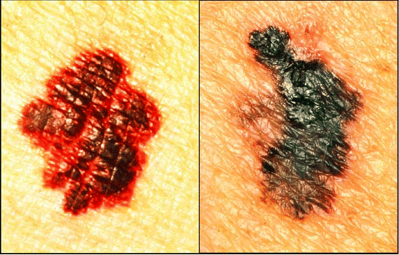
Sentinel node biopsy has proven its value in a number of cancers, including in clinically node-negative melanoma of the head and neck region. However, experts agree that it remains unclear what role sentinel node biopsy should have in other head and neck cancers. Clinical trials are under way to clarify the issue.
Sentinel Node Biopsy in Melanoma
Sentinel node biopsy was initially developed by Donald L. Morton, MD, Chief of the Melanoma Program and Medical Director and Surgeon-in-Chief of the John Wayne Cancer Institute in Santa Monica, CA, and colleagues in the early 1990s. The goal of the procedure is to improve staging of clinically node-negative patients by identifying small metastases in the first lymph node or nodes to which the tumor drains. For tumor types, such as melanoma, that metastasize via the lymphatic system, a negative sentinel node should indicate that the tumor has not spread beyond the initial tumor bed and that all nodes in the nearby basin will be negative. By more effectively identifying patients who have early nodal metastases, the procedure also reduces the number of patients who have to undergo a complete lymphadenectomy.
Results from a large multinational randomized trial indicate that sentinel node biopsy prolongs disease-free survival in patients with intermediate-thickness clinically node-negative melanoma. In the trial, 769 patients had standard surgical resection of the primary tumor and a sentinel node biopsy. If the biopsy showed evidence of disease, the patient had a complete nodal dissection. In the control arm, 500 patients underwent standard tumor resection and regional nodal observation. If a patient subsequently developed a palpable or radiologically detectable nodal metastasis, he or she had a complete lymphadenectomy.
At five years, 78.3% of the patients in the sentinel node biopsy arm were disease-free compared with 73.1% in the observation arm. Of the patients who had been found to have positive nodes-16% in the biopsy arm and 15.6% in the observation arm-there was an overall survival advantage for patients treated in the sentinel node arm, with a five-year survival rate of 90.2% vs 72.3% in the observation arm. There was, however, no difference in melanoma-specific survival or overall survival between the two trial arms when all patients, both those with and without nodal disease, were included in the analyses. The current analysis is the third of five scheduled interim analyses, and investigators hope that an overall survival difference might appear in subsequent analyses.
In reality, looking at overall survival characteristics of the two populations is not the right question, said Dr. Morton, who led the trial. The right question is, ‘In the patients who have nodal metastases, does identifying them by sentinel node biopsy and removing them early have a therapeutic advantage over just observing them and removing them when they become clinically evident?’
In my opinion, for the subset of patients who have nodal metastases, it is very clear that removing them early provides a survival advantage, as opposed to observing them until you can observe them by palpation, he said. That opinion is borne out by the disease-free survival advantage in the trial.
Additionally, having a negative sentinel node biopsy can provide patients with improved quality of life in several ways. The surgery is less extensive than a complete node dissection and is associated with less morbidity. The peace of mind that comes from having-in experienced hands-a negative sentinel node biopsy is huge, added Dr. Morton.

Given the level of evidence from this trial, the procedure should be used more consistently in the United States and around the world, said Charles M. Balch, MD, Professor of Surgery and Oncology at The Sidney Kimmel Comprehensive Cancer Center at Johns Hopkins University in Baltimore, who wrote the New England Journal of Medicine editorial that accompanied publication of the trial. This will be important for staging melanoma for people who have enough risk of metastases, he said. Patients with a thin melanoma have little chance of metastases, but for patients with melanomas that are of intermediate thickness, are thin with ulceration, or penetrate the dermis, the risk jumps to a 10% to 15% risk of spread.
Already it is being done routinely in most centers, though not in all centers, Dr. Balch said. It is currently included in the National Comprehensive Cancer Network treatment guidelines for intermediate-thickness melanomas.
Dr. Morton and the other investigators involved in the randomized trial have initiated a second randomized trial to compare the therapeutic value of sentinel node biopsy to complete node dissection in patients who have evidence of nodal metastases by sentinel node detection. The trial started in 2005 and is currently accruing patients.
The Procedure
Despite the acceptance of sentinel node biopsy as a standard of care for melanoma staging, the procedure is not likely to be offered at every community hospital in the United States in the future because it is technically demanding and its success depends directly on the surgeon’s skill, particularly for head and neck cancers. The learning curve is steep and requires 30 to 50 surgeries for a surgeon to become proficient, Dr. Morton and others agree. In the recent international trial, surgeons were required to have performed 30 sentinel node biopsies before they could enroll patients. Even so, the false-negative rate in the trial was 3.4%, whereas Dr. Morton’s own false-negative rate is 1.5%. We have shown in another paper that it is purely experience-driven. The greater the volume of patients a center has, the lower their false-negative rate, Dr. Morton said.
To identify a sentinel node, the surgeon injects the site of the primary tumor with a mixture of blue dye-often isosulfan blue-and a radioactive isotope, usually technetium-99m bound to sulfur colloid. The surgeon and a nuclear medicine specialist then use a handheld gamma counter to identify the hot spot on the skin; if lymphoscintigraphy is used to image the region, the lymphatic channel can often be identified. Once the surgeon has localized the sentinel node or nodes on the skin, he or she can open up the region, bearing in mind that if the node is positive the surgeon will likely use the same incision site to remove the remaining nodes. The lymphatic channel and sentinel node will appear blue due to the presence of the dye. To ensure that all the sentinel nodes have been identified, the removed nodes should be held up to a gamma counter to ensure that the majority of the radiation is accounted for. Surgeons will often remove any additional nodes that contain as much as 10% of the radioactivity in the hottest node.
Once the sentinel node is removed it can be sectioned and stained with standard H&E staining and with immunohistochemistry for disease-specific markers. Because the pathologist is examining only a few nodes rather than all of the nodes in a basin, the nodes can be analyzed more intensively, and smaller micrometastases identified if present.
Unlike the axilla or groin, which have relatively simple lymphatic drainage patterns, the neck region is complex. To illustrate that point, Dr. Morton noted that although surgeons were able to identify sentinel nodes in 99% of the axilla and groin lymph basins in the trial, they succeeded in finding sentinel nodes in only 84% of basins in the neck. Moreover, whereas 70% of the time there are only one or two sentinel nodes in the axilla and groin, the neck region frequently has more than two. I’ve seen up to six sentinel nodes, Dr. Morton said. Because the drainage is so complex, there are more likely to be multiple sentinel nodes, and therefore surgeons are more likely to miss one.
(A video of sentinel node biopsy for melanoma is available as part of the supplementary material included with the publication of the randomized controlled melanoma trial. The video can be found at http://content.nejm.org /cgi/content/full/355/13/1307/DC1, or by going to the New England Journal of Medicine Web site.)
Sentinel Node Biopsies in Head and Neck Cancers
The use of sentinel node biopsy in nonmelanoma head and neck cancers is still under discussion. Here is the problem, said Jonas T. Johnson, MD, Professor and Chairman of the Department of Otolaryngology and Professor of Radiation Oncology at the University of Pittsburgh School of Medicine. In melanoma, where it has been validated as actually affecting outcome, they have been able to study it in thousands of patients. In head and neck cancer, it has only been studied in hundreds of patients. Preliminary evidence suggests that it may be useful.
It is unequivocally true that about 70% of people undergoing complete neck dissection are found to have no nodal metastases, so in retrospect you could say they didn’t need it. But you didn’t know that until it was done. And sentinel node biopsy is not sensitive enough yet to replace that, in my opinion, he said.
To determine just how sensitive the diagnostic procedure is in squamous cell head and neck cancer, the American College of Surgeons Oncology Group (ACOSOG) designed a single-arm trial to compare the rate of nodal metastases detected by sentinel node detection and by subsequent selective removal of level I, II, III, and IV nodes. Complete data have not been released from the trial, but Francisco J. Civantos, MD, Associate Professor of Head and Neck Surgery at the Miller School of Medicine at the University of Miami, who led the study, presented preliminary data at the Triological Society meeting in February. A total of 137 patients with T1 or T2 clinically node-negative oral cancers were enrolled at 25 institutions by 34 certified surgeons. Preliminary data indicated a 92% negative predictive value with the technique, meaning that 11 of the patients had a negative sentinel node biopsy but were found to have nodal metastases upon more extensive nodal resection.
Tumors on the floor of the mouth appear to be more problematic for sentinel node biopsy than do those on the tongue or elsewhere in the oral cavity. One problem is that such tumors can lie right on top of the nodal basin, which makes distinguishing sentinel nodes difficult during surgery due to radioactive shine from the primary injection site. Additionally, the lymphatic drainage is particularly complex in this area, said Randal S. Weber, MD, Chairman of the Department of Head and Neck Surgery and Professor of Surgery at the University of Texas M. D. Anderson Cancer Center in Houston. The floor of the mouth has a high incidence of cross-lymphatics, which drain to the opposite side, for example. Based on these observations, Dr. Weber is cautious about the use of sentinel node biopsy in squamous cell head and neck cancer. It doesn’t appear to produce the same consistency of findings as it does in cutaneous melanoma.
Even with the strong preliminary data from the ACOSOG trial, researchers caution that a trial testing the efficacy of sentinel node biopsy alone, without complete neck dissection, would be needed before the procedure should be used as the standard of care. Such a trial could be a randomized trial similar to the one Dr. Morton and colleagues recently completed, in which patients were randomly assigned to either sentinel node biopsy or observation of the nodal basin, with lymphadenectomy if positive nodes are detected.
Not everyone is waiting for such clinical trial data, however. For example, Barry M. Rasgon, MD, Attending Physician and Director of the Research Otolaryngology Head and Neck Residency Program at Kaiser Hospital in Oakland, CA, has been using the procedure for about 10 years, in both melanoma and squamous cell cancers of the head and neck. The thing about sentinel node biopsy is that it is, bar none, the most accurate, cost-effective, least invasive way to stage a clinically negative neck.
He admitted that there are numerous potential pitfalls with the procedure but thinks they can be overcome without too much difficulty. When he first started trying the technique, he followed the general surgery literature in terms of the amount of radioactive tracer and volume to inject. It was a big black hole. Everything was hot, he said. With a substantial reduction in both radioactivity and volume, to about 30 microCuries in 0.1 or 0.2 mL, the nodes become apparent, and the shine from the primary injection site does not overwhelm everything else. To further minimize the problem when he is dealing with a squamous cell cancer on the tongue, Dr. Rasgon bends a small piece of lead around the primary injection site. Even with such hints, though, it can be a tough technique to master because of the complex lymphatic drainage in the neck. You probably need to do about 30 of these with someone who knows what they are doing to get good at it, Dr. Rasgon said.
Remarkably, the technique has enabled him to identify two recent cases in which micrometastases in the sentinel nodes had spread beyond the node itself. We think there needs to be a certain volume of tumor in the node to do that. But clearly it can do that from a small focus of micrometastases in the paracortical area and go right through the capsule to the surrounding tissues, Dr. Rasgon said.
Nonetheless, sentinel node biopsy is still considered an experimental procedure by most clinicians. My advice is that patients with squamous cancer of the upper aerodigestive tract, who are staged N0, benefit from having elective selective neck dissection based on anatomical risk, Dr. Johnson said. I think sentinel node biopsy for aerodigestive cancer is not yet ready. It is not yet the standard of care.
©2007 The Triological Society


Leave a Reply