Revision endoscopic sinus surgery (RESS) has challenges that often are not seen in primary surgeries. Still, there are tips to keep in mind when working with issues such as altered anatomy, a lateralized middle turbinate or return polyp patients.
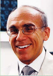
To address these issues, ENToday asked two leading surgeons for their take on RESS. Michael Friedman, MD, Professor of Otolaryngology at Rush University Medical Center in Chicago, and Raj Sindwani, MD, Assistant Professor of Otolaryngology at St. Louis University, discussed which patients benefit the most from RESS, when it is indicated, its limitations, and what the future holds.
Indications for RESS
Generally, RESS is part of the treatment for chronic sinusitis, said Dr. Friedman. While there are data on the number of people with chronic sinusitis (30 million in the United States), and the number of sinus surgeries performed in general (about 300,000 annually), no one knows exactly how many of those surgeries are revisions-estimates in the medical literature vary from 20% to 60%.
Chronic sinusitis is the most common chronic condition in the United States, Dr. Sindwani said. Studies show that the quality of life of patients with chronic sinusitis is worse than COPD, asthma, and even things like chronic back pain, he said. In his own tertiary level practice, revision cases outnumber primary cases.
There are two types of patients who return for sinus surgery: the polyp patient, and those with no polyps. Patients who have polyps are challenging because of the high recurrence rate. In fact, when patients go in for primary polyp surgery, it’s a good idea to discuss the fact that the disease is likely to recur and that further surgeries will be needed. That way, when second, third, or fourth surgeries are needed for polyps, previous surgeries will look less like failures in the eyes of the patient, said Dr. Friedman.
Patients who have polyps have an intrinsic disease of their sinus lining. They almost always have some sort of allergy problem and a significant percentage have an allergic fungal sinusitis…. Surgery can’t correct the underlying problem, Dr. Friedman said. Returning polyp patients become surgical candidates only after a rigorous arsenal of treatments to fight the disease has had an insufficient effect.
In some ways, repeat surgeries for polyp patients aren’t true revisions, said Dr. Sindwani. You’re not really revising per se, you’re going back in because new polyps have grown, he said. With polyp patients, going back in sometime in the future is part of the overall treatment plan-unlike other types of revision surgeries.
Even though some patients need multiple surgeries for polyps, most studies looking at patient satisfaction show it’s worth it.
But some patients who return may have additional sinus troubles. There could be scar tissue from previous surgeries, residual infected tissue (which can cause recurrent polyps), or tissue that didn’t heal properly. In addition, primary surgery may have caused the middle turbinate to scar or even lateralize blocking the sinus opening, for example.
Most of these issues are common reasons for post-sinus surgery trouble even in non-polyp patients. The middle turbinate can be a tricky issue, however.
Dealing with the Middle Turbinate
Historically, surgeons would remove the middle turbinate because it could block the sinus opening. A complication of sinus surgery, in general, is that the middle turbinate lateralizes to a position where it scars or blocks airflow into the frontal sinus, maxillary sinus, or the anterior ethmoids. Even in the best conditions, it can happen, said Dr. Friedman.
Although some surgeons opt to resect the middle turbinate to avoid this, most sinus experts now opt to preserve the natural anatomy. If it scars, the problem would be evident in an early postoperative visit and at this point an early revision could be done. Some revisions are required as early as one or two months after surgery, he said.
Revision Technically More Difficult than Primary Surgery
When it comes to revision surgery in the non-polyp patient, a big challenge is working with anatomy that has been changed. If a patient has undergone sinus surgery, and if the first surgeon did an incomplete job in terms of removing tissue, then the second surgical procedure is a completion of the first surgery, Dr. Friedman said.
Revision surgery is more difficult because the anatomy has been distorted, it is technically challenging, and the risk of complications is higher.
For surgeons doing primary procedures, approaching the sinus with the intent to treat all the affected areas is important, he said. Some surgeons may be comfortable with operating on just the ethmoid and maxillary sinuses, and aren’t comfortable with the frontal or sphenoid sinuses, he continued.
If a patient needs more extensive surgery, refer him or her to a more experienced surgeon. Doing only part of it can be worse than doing nothing, Dr. Friedman said.
Dr. Sindwani agreed that revisions can be fraught with difficulties. Even if you did do the primary surgery, there can be infection, scarring, and inflammation, making it easy to become disoriented during RESS. The anatomic landmarks that usually guide a surgeon through the sinuses are often distorted or may be absent altogether, he said.
In polyp patients, distorted anatomy, loss of landmarks and potential surgical disorientation are of even greater concern, as it is often in the setting of extensive disease and increased bleeding as well, Dr. Sindwani said.
Image-Guided Surgery
Revision surgery is a good reason to use an image-guided system. CT helps with preoperative planning for revision surgery. Intraoperatively, these powerful systems will localize the position of the surgeon’s instrument in relation to the patient’s sinus anatomy in a real-time fashion, and thereby improve the surgeon’s ability to stay oriented and safe, Dr. Sindwani said.
A few companies have come up with less expensive, more compact image-guidance systems that have made it possible for surgeons to perform image-guided RESS in settings outside of large hospitals and major academic centers, he said.
Image-guided technology is especially important for the frontal and sphenoid sinuses, Dr. Friedman said.
Another point about CT is that what is looked for in revision cases is different from what is looked for in primary cases, he said. The CT for revision cases is often less dramatic than in primary cases who have not had surgery to help with clearing of secretions. There is less opacity.
The revised patient may have had surgery to open a passageway, but if a new passageway was created and the natural one is still blocked, the patient may still be getting infections along with symptoms of purulent secretions.
It is key to use endoscopic findings to clarify what is happening, Dr. Friedman said.
Postoperative Care
Good postoperative care can reduce the need for RESS, Dr. Sindwani said. Seeing a patient in the office one or two weeks after surgery, once or twice, to inspect how well the sinuses are healing is good practice.
If there is evidence of early scarring or synechiae formation in critical areas, this can be dealt with in the office while the scars are still immature. You can take down some scarring in the clinic using topical anaesthesia, he said.
The middle turbinate should be watched. In cases where he has had to work on sinuses where the middle turbinate had been lateralized after previous surgery, Dr. Sindwani said he can take steps to ensure that the same thing does not happen again. This can include total or partial resection of the turbinate, the use of spacers, or using endoscopically placed sutures to hold the turbinate in place (against the septum) during the RESS in the OR.
If the turbinate is noted to have shifted during a postop checkup, it can be gently nudged back in place with the placement of a spacer to hold it over while it heals, Dr. Sindwani said.
Future Look
Generally, the frontal sinus is the toughest to keep open and is commonly associated with recurrent trouble. In stubborn cases, stents are sometimes used. However, there is controversy as to how long they should stay in and they can be associated with their own sets of problems, Dr. Sindawani said.
Dr. Friedman agreed that stents are tricky. On the one hand they prevent scarring or restenosis. But it’s a foreign body and the stent itself can become obstructed with mucus, he said.
An alternative now being studied in Dr. Friedman’s center is the use of balloon sinusplasty for patients with postoperative scarring. A microdebrider is used to clear some space, then with the use of fluorososcopy, a small wire is threaded into place. The balloon is placed over the wire, then inflated under high pressure. The ostium is stretched open instead of being cut, he said.
Initial clinical studies show that the technique is easy, with low morbidity. I think this is something for the future for patients with frontal sinus scarring and sphenoid sinus scarring, and possibly, to a lesser extent, maxillary sinus scarring, he said.
Dr. Sindwani cautioned that balloon sinuplasty has not been studied rigorously enough to endorse yet. When it comes to keeping the frontal sinus open, which indeed can be tough. Balloons used to open this area more than likely is enlarging the frontal recess (the area just below the frontal opening), rather than the true opening itself, he said.
Overall, the best way to avoid RESS is to use meticulous surgical technique and avoid inadvertent tissue trauma in critical and unforgiving areas like the frontal and sphenoid sinuses, Dr. Sindwani said.
©2007 The Triological Society

Leave a Reply