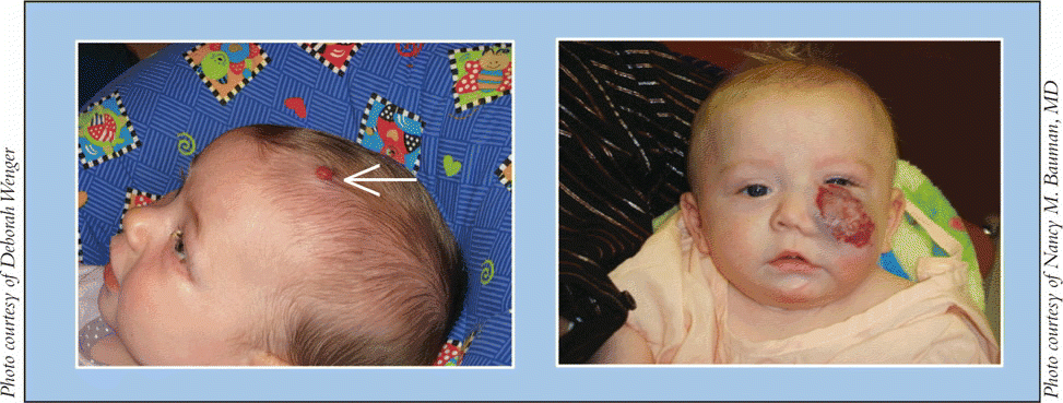
Infantile hemangiomas and lymphatic malformations (LM) are vascular anomalies that otolaryngologists-head and neck surgeons often encounter in their practices. Infantile hemangiomas and LMs differ from one another in prevalence, etiology, and clinical presentation, but both may be undergoing potential shifts in treatment, depending on research outcomes.
Explore This Issue
June 2009Appropriate classification of vascular anomalies is integral to their successful management, said Nancy M. Bauman, MD, Professor of Otolaryngology-Head and Neck Surgery at Children’s National Medical Center in Washington, DC.
The International Society for the Study of Vascular Anomalies (ISSVA) adopted the classification scheme proposed by John Mulliken and Julie Glowacki in 1996, recognizing vascular tumors as lesions with endothelial cell hyperplasia, or hemangiomas, and vascular malformations as lesions arising due to dysmorphogenesis, or LMs (see table).
Hemangiomas
Infantile hemangiomas are the most common tumor of infancy and arise in 5% to 10% of infants, with two-thirds of lesions involving the head and neck, said Dr. Bauman. Hemangiomas affect females about three times as often as males, and arise more commonly in Caucasians and preterm infants.
Proposed etiologies of hemangiomas include defects in angiogenesis, mutations in growth regulatory genes of endothelial cells, and embolization of placental microvessels, said Dr. Bauman.
Tumors first become clinically apparent when a child is two to four weeks old, said Elaine Siegfried, MD, Professor of Dermatology at St. Louis University. You typically don’t see them at birth, she said. They usually grow for about six to 12 months and then take two to 10 years to involute, she explained.
We once believed that asymptomatic infantile hemangiomas should not be treated, since they will eventually involute, said Dr. Bauman. We now know that some of these hemangiomas-particularly large lesions of the lip or the tip of the nose-will cause permanent disfigurement, and early intervention may improve the cosmetic outcome before the lesions permanently affect the underlying structures.

Complications
Textbooks generally don’t describe different types of hemangiomas, but physicians recognize that subsets of high-risk and low-risk tumors exist, said Dr. Siegfried. High-risk hemangiomas tend to be segmental, while low-risk tumors are generally focal, she noted.
Most hemangiomas are relatively asymptomatic and should be allowed to involute on their own, but about 25% produce symptoms related to their size and location, noted Dr. Bauman. For example, periorbital hemangiomas can obstruct the visual axis and affect vision development, she said.
Leave a Reply