Microdebrider surgery, a technology with its roots in the rotary vacuum shaver introduced by Urban in 1968 for removal of acoustic neuromas, is now being successfully utilized for many different types of procedures. The microdebrider has been used for several years by orthopedists for joint surgeries. Plastic surgeons are employing its use for liposuction. More recent research has shown the microdebrider to be an effective tool for several types of otolaryngologic procedures, including, but not limited to, sinus surgery and tonsillectomy.
What Is a Microdebrider?
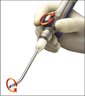
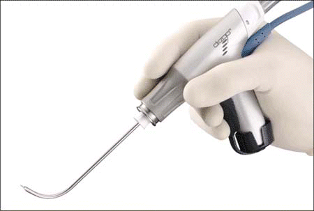
The microdebrider is a tool consisting of three key components. The console, controlled by a foot pedal, determines the speed and direction of the rotating blade. Blades can be rotated in forward, reverse, or oscillating modes at various speeds.
The handpiece controls the blade and integrates suction to allow for rapid and simultaneous removal of debris. It is compatible with blades of various sizes and configurations. Suction tubing connects to the handpiece.
The blade (or bit) is a hollow metal tube with a port for suction. Blades can be smooth or serrated and are available in various sizes. This design allows for simultaneous cutting and removal of tissue. The device is designed for one-time use and is disposable.
Because of the diameter of the blades (5-7 mm) and the rigidity of the microdebrider, it cannot be used with a flexible scope; it must be used in conjunction with a rigid bronchoscope or suspension laryngoscope.1
How It Works
The microdebrider works by employing suction to pull tissue into the aperture of the blade, which cuts the tissue. Suction is used to simultaneously remove tissue-and blood-from the site, allowing much better visibility for the surgeon. The tool can be easily operated with just one hand, allowing the operator more freedom of movement.
The only true risk identified by investigators involved with the use of the microdebrider is the possibility of inadvertent resection of normal tissue, and potential injury to the patient as a result. This most commonly occurs when a drilling bur is used, but it can occur with cutting blades as well. This risk can be virtually eliminated if the operator remembers to:
- Avoid overzealous suction pressure (which can pull excessive tissue into the blade).
- Always maintain adequate visualization of the blade or bur.2
Microdebridement of Obstructing Airway Lesions
Tracheobronchial obstruction will occur in an estimated 20% to 40% of lung cancer patients. Airway obstruction can also occur from certain benign conditions such as postintubation tracheal stenosis, relapsing polychondritis, and Wegener’s granulomatosis. The obvious goal of debridement of an obstructing airway lesion in an acute situation is to restore oxygenation to the patient. With the added risks associated with general anesthesia, the ideal intervention will be fast, effective, and carried out in a manner that poses the least risk of additional injury to the patient.
Removal of the lesion may be diagnostic as well as palliative, so preservation of tissue for pathological review may be desirable. In cases of malignancy, the debridement of an obstructing lesion can often buy extra time for the patient’s oncologic team to put together a comprehensive plan for oncologic, psychologic, and nutritional management before beginning definitive treatment.3
William Lunn, MD, is Director of Interventional Pulmonology and Assistant Professor of Medicine and Otolaryngology at Baylor College of Medicine. Dr. Lunn was primary author of a study published in The Annals of Thoracic Surgery in September 2005, titled Microdebrider Bronchoscopy: A New Tool for the Interventional Bronchoscopist. Dr. Lunn stated in a telephone interview that he saw a fellow otolaryngologist use the microdebrider for sinus surgery in about 1998, and he and his colleagues thought this would be a great tool to use in the airway. In this study, the authors reported their experience using the microdebrider to treat central airway obstruction.

Dr. Lunn et al. reported that obstructing airway lesions were rapidly removed using the microdebrider in all 23 patients included in the study. Only mild bleeding occurred, even in lesions that appeared to be quite friable and vascular. Bleeding that did occur was controlled by tamponade using the rigid scope or instillation of oxymetazoline hydrochloride. All patients concluded the procedure free of ventilatory support. No further intervention for airway obstruction was required in follow-up ranging up to 24 months following the procedure.
When thermal modalities are used for treatment of obstructing airway lesions, one of the primary dangers is the possibility of an airway fire. Interventions can be quite prolonged because the surgeon must debride, suction, and cauterize sequentially. There is also the potential for causing injury to the airway or surrounding structures, which may require further interventions to correct.
According to Dr. Lunn, the traditional methods of debridement of airway lesions require three instruments in the airway: a telescope, forceps or a cutting device, and suction. Using the microdebrider, the surgeon can hold the debrider in his or her dominant hand, and the other hand is free to hold the scope.
The advantages of microdebrider surgery for airway obstruction cited in The Annals of Thoracic Surgery study include:
- The ability to rapidly remove obstructing tissue.
- The ability to simultaneously remove tissue debris and blood allowing for better visualization of the field.
- The ability to precisely limit the effects of the modality without fear of combustion or perforation of the airway.
Another advantage cited by Dr. Lunn is that the rigidity of the tool allows for a tactile feel of the airway wall. It is possible to simply stop the rotation of the blade and use the blade to touch and feel the airway, something that can’t be done with the filament of a laser.
When asked if there are cases of airway obstruction in which use of the microdebrider might be contraindicated, Dr. Lunn responded, No. There’s really nothing. If I need to remove the tissue anyway, I think the debrider is the most delicate way to do it.
Partial Tonsillectomy
Tonsillectomies are performed on more than 300,000 patients annually in the United States, primarily for chronic obstruction or infection. In traditional tonsillectomy, the removal of the capsule enclosing the tonsil leaves the throat muscles, large blood vessels, and nerves exposed. Bleeding is generally controlled by electrocautery. This process leaves the patient with severe pain and swelling, frequently requiring the patient to take pain medications for several days. This pain and swelling can also lead to decreased oral intake, causing dehydration that may in some cases even require further hospitalization. These effects can significantly delay the patient’s return to normal activity.
In a study published in Otolaryngology-Head and Neck Surgery in January 2005, researchers concluded that use of a microdebrider for partial tonsillectomy resulted in less pain for patients and shorter recovery times. The study, supported by an unrestricted grant from Medtronic, introduced the powered intracapsular tonsillectomy and adenoidectomy (PITA) technique utilizing a microdebrider tool. Using this technique, physicians removed 90% to 95% of the tonsils, leaving the capsule in place to protect the throat muscles, blood vessels, and nerves.
In this study, patients’ caretakers were asked to record pain levels, doses of pain medications, time of return to normal activity, and time of return to normal diet following tonsillectomy. Patients who had partial tonsillectomy utilizing the microdebrider were three times more likely to not require pain medicine three days following surgery, and almost twice as likely to have resumed normal activity levels than patients receiving traditional tonsillectomy.4
Sinus Surgery
Sinusitis can have a significant impact on quality of life and is an extremely common health problem in the United States. An estimated 37 million Americans suffer sinusitis attacks lasting for at least 12 weeks or recurring frequently. Although most individuals who suffer from sinusitis can be treated with medications, about 10% of sufferers require surgery to obtain relief. Sinus surgery has a long-standing reputation of being especially brutal for patients, requiring external incisions and nasal packing, and leaving patients with pain and bruising for weeks following the procedure. Use of the endoscope has made sinus surgery much less traumatic, eliminating the need for external incisions. Use of the microdebrider can allow for even more delicate tissue removal, reducing the need for separate grasping and cutting instruments.5
Steven Pletcher, MD, Assistant Professor in the Department of Otolaryngology-Head and Neck Surgery, Division of Rhinology, at the University of California, said that the advantage of the microdebrider is that cutting and suctioning can be accomplished simultaneously with one hand. This can be helpful when performing sinus surgery, when, as Dr. Pletcher said, You’re operating in a deep cavity and you have room for only one single-handed instrument.
Microdebrider Eustachian Tuboplasty
Dr. Pletcher coauthored a study presented at the American Academy of Otolaryngology-Head and Neck Surgery meeting titled Microdebrider Eustachian Tuboplasty: A Preliminary Report. In this study, the microdebrider was used to treat patients with eustachian tube dysfunction. The patients involved in the study all had concurrent sinonasal disease and underwent endoscopic sinus surgery at the time of microdebrider eustachian tuboplasty. The authors concluded that the procedure is safe and may provide relief from symptoms of eustachian tube dysfunction. The researchers reported no surgical complications, and subjective symptoms of ear blockage improved in 70% of patients included in the study.

Dr. Pletcher concluded, We did get good results, but the patients also had sinus surgery at the same time. We didn’t have the ability to determine if it was the sinus surgery or the procedure that involved the eustachian tube specifically that made the symptoms resolve. He and his coauthors determined that further studies will be needed to make this determination.
As with others researching use of the microdebrider, Dr. Pletcher also cautions that because of the rapid removal of tissue, surgeons must be very careful about the tissue they are targeting for removal. He states that the microdebrider can remove a great deal of tissue very quickly, so the operator must be very cognizant of the location of the blade at all times and maintain a healthy respect for the risks.
Cost
Because the microdebrider has disposable parts, it can be more expensive than other surgical tools. However, Dr. Pletcher said that he feels he can operate more quickly using the microdebrider. He opined that whatever may be lost in the cost of the device itself is made up and possibly saved in time.
Dr. Pletcher has encountered no difficulties with insurance coverage of the microdebrider as an instrument in association with other procedure costs. He concluded, The cost issue is probably negligible.
The Future of the Technology
According to Dr. Lunn, manufacturers plan to combine cautery with the debrider. Suppliers are working on developing debrider blades with cautery function, and at least one is already in production. He stated that the development of such a combination tool would be even more time-efficient, eliminating the need to switch tools when cauterization is required. He speculated, That would be really neat for the debriders. The device would have three functions: cut, suction, and cauterize.
Dr. Lunn also reported that at least one company has recently started marketing longer blades, which make it possible to reach deeper lesions. He said that with these longer blades it is now possible to go all the way down the length of the mainstem bronchus into the segmental bronchi.
Studies have shown the microdebrider to be an effective surgical tool for many widely diverse types of procedures. Researchers have deemed it a convenient and efficient tool, with many advantages and only one identified risk, albeit a potentially significant risk.
Dr. Pletcher summed up his opinion based on his experience: I think the most important point with the microdebrider in terms of sinus surgery is that it’s one of the most effective tools, but potentially one of the most dangerous tools. The debrider needs to be used with the appropriate respect for complications that can result from its use.
References
- Lunn W, Ernst A. The microdebrider allows for rapid removal of obstructing airway lesions. CTSNet. Available at www.ctsnet.org/sections/thoracic/newtechnology/article-6.html . Accessed 12/5/06.
- Lunn W, Garland R, Ashiku S, et al. Microdebrider bronchoscopy: a new tool for the interventional bronchoscopist. Ann Thorac Surg 2005;80:1485-88.
- Simoni P, Peters GE, Magnuson JS. Use of the endoscopic midrodebrider in the management of airway obstruction from laryngotracheal carcinoma. Ann Otol Rhinol Laryngol 2003 Jan;112(1):11-13.
- Medtronic news release 2/5/05. Available at www.medtronic.com/Newsroom/NewsReleaseDetails.do?itemId=1139418429133⟨=en_US . Accessed 12/5/06.
- Saltus R. Breathing easier breakthrough surgery takes the agony out of repairing blocked sinuses. The Boston Globe. 12/5/2000. Available at www.boston.com . Accessed 1/1/07.
©2007 The Triological Society


Leave a Reply