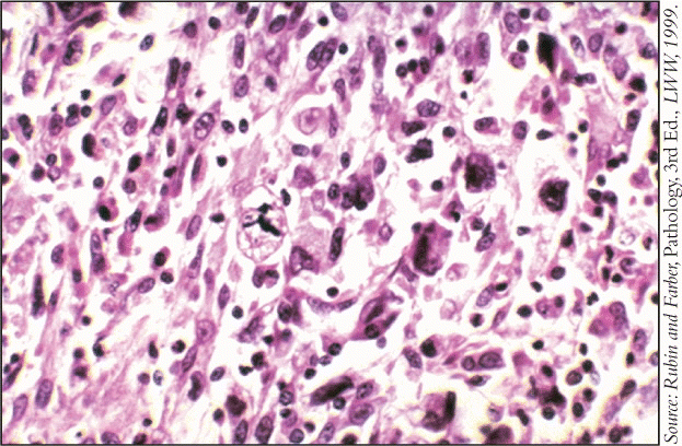
Patients also may have an elevated eosinophil count, hypoglycemia, fever, and abnormal liver function tests.
Explore This Issue
May 2009The only definitive diagnosis is biopsy, but imaging studies are usually done first.
- MRI provides good contrast between tumor and muscle, excellent definition of the surrounding anatomy, and ease of imaging in multiple planes. It reveals an intramuscular mass with heterogeneous signal intensity on all pulse sequences.
- CT scan is preferred by some, but the definition between tumor and muscle is not as good. It typically shows a large, nonspecific, lobulated soft tissue mass with density close to that of muscle.
- Ultrasonography shows a well-defined heterogeneous mass, but the appearance of tumors on ultrasound is nonspecific. It can, however, facilitate directed needle biopsy.
Biopsy is all-important-and not at all straightforward. For accessible lesions of the upper aerodigestive tract, paranasal sinuses, or scalp, direct biopsy is appropriate. For inaccessible lesions, such as those in the neck, fine needle biopsy should be the first step. Core needle biopsy can usually definitively diagnose sarcoma, and often allows subtyping. If incisional or excisional biopsy is necessary, the surgeon must consider the approach carefully.
Treatment
Assuming a correct diagnosis, early and complete surgical excision is the best initial treatment. The higher the stage of the disease, the wider the margins must be. For larger tumors, preoperative radiation and/or chemotherapy can shrink the tumor.
In the head and neck, there are many vital structures in a confined area, said Dr. Clark. The classic mantra regarding margins is to resect to a new tissue plane, but with limited space and vital structures, this presents a challenge. Therefore, surgery for larger lesions, those of the skull base, and those adjacent to vital structures, such as the eye, can be quite morbid.
Patients with MFH often have had more than one episode before diagnosis, so the tumor is only partially resected, thus increasing the potential for recurrence and/or distant metastasis.
The usual chemotherapy agents include ifosfamide, high-dose ifosfamide with mesna to prevent hemorrhagic cystitis, cyclophosphamide, doxorubicin, vincristine, adriamycin, gemcitabine, and taxanes. Combination chemotherapy is almost always more effective then use of a single agent.
No one agent has emerged as the treatment of choice, said Dr. Clark. The rarity of the tumor precludes sufficient enrollment in controlled studies, which continues to be a significant limitation.

Leave a Reply