At first glance, the Otolaryngology Surgery Simulation Center at Montefiore Medical Center in New York resembles a traditional temporal bone dissection lab. Two sets of stations face each other, their T-bone samples in jars waiting for third-year surgical residents to try mastering complex and delicate surgery. But something is different. The stations are flanked on either side by two medical simulators-one for endoscopic sinus surgery (ESS), the other for temporal bone dissection (TBD). The temporal bone stations are otolaryngology’s past; the simulators are its future.
Babak Sadoughi, MD, of Montefiore’s simulation center, explained: Using cadaveric bones presents many issues. Drilling human bones is messy, it’s hard to find cadavers, and there are contamination issues and strict disposal protocols to follow. Also, once you drill a bone, you’re done.
With the TBD simulators, Montefiore’s 20 otolaryngologic surgical residents can practice T-bone dissection to their hearts’ content. Montefiore’s Chair of Otorhinolaryngology-Head and Neck Surgery, Marvin Fried, MD, a pioneer in using otolaryngologic simulation since the 1980s, inherited the ESS simulator when he came to Montefiore, then figured out how best to use it.
 It’s a multidisciplinary effort including otolaryngologists, neurosurgeons, CT and MRI experts, physicists, biomedical engineers, and computer experts to develop these standards to the level where they could impact otolaryngology and surgical training.
-Gregory Wiet, MD
Medical simulation can change everything but it still has a long way to go, said Dr. Fried. There are significant funding shortfalls interfering with our ability to perform validation studies that would eventually lead to use in board certification. That’s years away. Patient safety requirements driven by AHRQ [the Agency for Healthcare Research and Quality] should help. After all, we operate in the same areas of the anatomy as neurosurgeons, so reducing errors is critical, he added.
The otolaryngologists-head and neck surgeons at Montefiore are part of a national movement to develop and integrate medical simulation into research, education, clinical practice, certification, and maintenance of credentials. Medical simulation, a subset of virtual reality, uses computer-generated images, video cameras, models, and sensors for learners to practice procedures in lifelike environments. It enhances learning with visual, auditory and haptic feedback to help physicians feel that they are making decisions and performing procedures in real life. (See part 1 of this series, ENT Today, July 2008, p. 1.) The work of some otolaryngology innovators is described below.

Cutting Edges
Gregory Wiet, MD, a pediatric otolaryngologist-head and neck surgeon and Associate Professor at Ohio State University, recognized simulation’s potential while working as a surgical resident at OSU’s Supercomputer Center in 1993-94. I was enamored of the pretty pictures, and the visualization of human anatomy. I realized that for otolaryngologist-head and neck surgeons and neurosurgeons, having a 3D reconstruction of a tumor on our laptops could help us in preoperative planning and surgery. That was novel thinking in 1993, but it’s still valid today. Dr. Wiet is deeply involved invalidating data from surgical simulations.
With Don Stredney, PhD, Director of OSU’s Supercomputer Center, Dr. Wiet obtained critical funding from several sources, especially the National Institutes of Health, to standardize CT and high-resolution MRI studies on which to build a T-bone dissection simulation. It’s a multidisciplinary effort including otolaryngologists, neurosurgeons, CT and MRI experts, physicists, biomedical engineers, and computer experts to develop these standards to the level where they could impact otolaryngology and surgical training, Dr. Wiet said.
Dr. Stredney said that getting funding for validation can be frustrating, but is critical for success. It’s too bad that we’re not on the video game industry’s radar, because they sell a million units a month, but what we’re doing doesn’t interest them.…The data sets we arrive at are the sine qua non of simulation. That’s what you need for an ENT surgical simulation, he concluded.
Not everyone is comfortable with simulation’s current stage of development. Bruce Gantz, MD, Professor of Otolaryngology at the University of Iowa and a specialist in cochlear implants and acoustic neuromas, has had his program’s surgical residents use TBD simulators. Our feedback is that they like it and it’s interesting, but not good enough to truly simulate surgery. It does not have the same ergonomics, the same feel as the real thing, he said.
Dr. Gantz has tried simulators from different vendors, but said he’d prefer his residents to spend their time with real temporal bones, which are in good supply in Iowa. Simulation is intriguing, but all of the systems I’ve tried have drag to them. Until it feels right in my hand, I’m not comfortable relying heavily on simulators to train residents, he added.
Trying to close the gap between simulation and the OR at Stanford University’s Center for Immersive and Simulation-Based Learning are Otolaryngology Associate Professor Nikolas Blevins, MD, and Kenneth Salisbury, PhD, head of the Bio-Robotics Laboratory and a scientific advisor to Intuitive Surgical. Their respective teams have targeted the physical interaction between human beings and computer-driven actuators to develop human-friendly robotics in simulation. Put simply, working on the feel of the simulator in the surgeon’s hand is a current interest. They also are reducing the amount of time surgeons have to spend on developing simulations by using simulator-generated algorithms and teaching tools. For example, in developing a mastoidectomy simulation, they had both novice and experienced surgeons perform virtual surgeries, and have collected data on drilling and suctioning techniques. They validate metrics by correlating the scores generated by algorithms versus seasoned surgeons’ ratings.
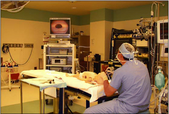
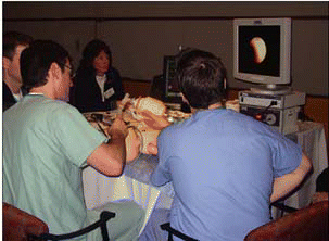
Ellen Deutsch, MD, a pediatric otolaryngologist at the Alfred I. duPont Hospital for Children, is excited about simulation’s providing opportunities for adult learners and for their teachers to be rejuvenated as they incorporate simulation in their curricula. Describing a simulation using a high-fidelity infant mannequin, Dr. Deutsch has the resident discern if the infant has aspirated a foreign body in the left main bronchus. The baby’s oxygen level is in the low 90s, with stridor, and only the right chest moves during respiration, indicating a possible obstruction. The learner must select the endoscopic equipment to expose the airway, perform endoscopy, see the foreign body, and remove it. The endoscopist must also have a conversation with the anesthesiologist. If the anesthesia is too light, the baby may experience laryngospasm, or if the endoscopy takes too long without the anesthesiologist ventilating the patient, the baby’s oxygen level could drop. The beauty of simulation is that the learner can do it over repeatedly, turning fumbles into competent fluency, said Dr. Deutsch.
 Using cadaveric bones presents many issues. Drilling human bones is messy, it’s hard to find cadavers, and there are contamination issues and strict disposal protocols to follow. Also, once you drill a bone, you’re done.
-Babak Sadoughi, MD
As an instructor, Dr. Deutsch works with a variety of professionals from different disciplines on developing and implementing various simulation projects, including in situ environments that heighten the learner’s experience of being in real clinical situations. Some of our ENT simulation practice opportunities are very real. In a sophisticated immersion environment, for example, we’re in a mock-up of a patient room, practicing on a high-fidelity infant mannequin with computer-controlled fontanels, vocal cords that can close, and dose-related responses to medication, said Dr. Deutsch.
The increasing variety of simulation tools that are now available allow users of medical simulation to select the most appropriate technology or strategy to accomplish specific objectives, and to provide an activated educational experience based on the individual needs of strategy based on the individual needs of adult learners.
Funding: An Obstacle to Progress
Whereas funding for ENT validation studies remains scarce, medical simulation’s cheerleaders find creative ways around the dollar drought. A number of major medical institutions with simulation centers spread the cost across various clinical, engineering, and IT departments (see www.medsimulation.com/education_partners/centers.asp for a list of centers). Developing a large institutional simulation infrastructure supported is cost-effective and encourages cross-discipline collaboration on new simulations.
Funding for validation studies remains otolaryngology’s stumbling block. Even after joining forces, Montefiore, NYU Medical Center, and New York Eye and Ear Infirmary cannot scrape together the approximately $15 million in funding required to validate simulations at the level required for inclusion in board certification. Support from the American College of Surgeons and the American Association of Neurological Surgeons hasn’t gotten them over the hump either. Governmental and private foundation funding sources have currently dried up.
Partnering with commercial vendors of otolaryngologic simulators has potential, but those vendors face a chicken-and-egg conundrum. They need committed customers before spending millions on developing sophisticated surgical simulations. Lockheed Martin Corporation, which, in the 1980s developed Martin, the first ESS simulator, exited the field after having built only four systems. Montefiore’s Dr. Fried, one of Martin’s first users, is now working with physicians and vendors associated with the VOXEL-MAN project, headquartered at the University Medical Center at Hamburg-Eppendorf, on finding a cost-effective way to build a new ESS simulator. We’re getting to the point where ENT simulations will be commercially viable. It may take five years but the potential is there. The stakes are high in both temporal bone and sinus surgery because of patient safety issues, he said.

Despite funding shortfalls, otolaryngologists-head and neck surgeons are embracing medical simulation. Students and teachers recognize it as a powerful new teaching tool, and surgical residents and experienced physicians are using it to hone or refresh their OR skills and prepare for delicate surgeries. As large hospitals and academic medical centers build their own simulation centers, they will draw on the expertise of their clinicians, administrators, social scientists, engineers, and IT departments to develop new tools and applications for medical simulation.
Medical Simulation Would Reduce Surgical Errors
- Screen surgical residents for demonstrable aptitude.
- Provide initial training in surgical experiences.
- Promote ongoing training of surgeons through consistently reproducible processes.
- Enable periodic assessment of acquired surgical skills.
- Maintain proficiency through rehearsal of complex, patient-specific procedures.
Source: Healy GB. The College should be instrumental in adapting simulators to education. Bull Am Coll Surg 2002;87:10.
©2008 The Triological Society
Leave a Reply