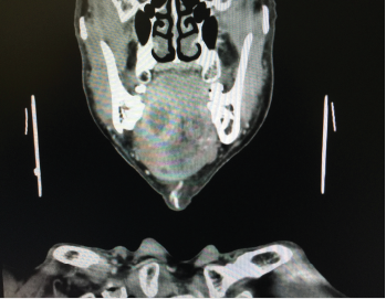
This CT scan shows a large abscess inside the tongue tissue.
©Karan Bunjean / shutterstock.com
It’s been nearly four years since the American Joint Committee on Cancer (AJCC) updated the staging system for oral cavity squamous cell carcinoma (OCSCC). Key revisions include factoring in depth of invasion (DOI) and extranodal extension (ENE) of the primary tumor (AJCC Cancer Staging Manual. 8th ed. Springer International Publishing: American Joint Commission on Cancer; 2017).
Explore This Issue
October 2021Although more work needs to be done to incorporate the AJCC8 staging system into clinical practice, it has already had an impact on more accurate staging of primary tumors, thus opening the door for better risk stratification and treatment, according to several head and neck cancer specialists.
The stakes for accurately staging OCSCC are high, given its high incidence and poor prognosis. The condition remains one of the most common cancers in the United States, affecting approximately 34,000 people each year. Five-year survival for patients with localized disease is approximately 75%; however, when the cancer has spread to the lymph nodes, survival rates drop to about 50%.
“We’ve all recognized for a while that the DOI of an oral cavity cancer is a very significant prognostic indicator, so this was a much-needed revision,” said Michael Moore, MD, Arilla Spence DeVault Professor of Otolaryngology–Head and Neck Surgery and medical director of the Indiana University Health Joe and Shelly Schwarz Cancer Center in Carmel. “By more accurately staging these cancers, we can better capture, from a prognosis standpoint, how these patients will do.”
Measuring Depth of Invasion
In the new AJCC staging system, the DOI is “measured first by finding the ‘horizon’ of the basement membrane of the adjacent squamous mucosa. A perpendicular ‘plumb line’ is established from the horizon to the deepest point of tumor invasion,” noted Cherie-Ann Nathan, MD, the Jack Pou Endowed Professor and chairman of the department of otolaryngology–head-neck surgery at Louisiana State University Health in Shreveport, La. Although studies have examined 3-, 4-, and 5-mm increments to determine lymph node metastasis and survival risk stratification, the experts involved in developing the AJCC8 settled on 5-mm increments, Dr. Nathan said (see Table 1).
By more accurately staging these cancers, we can better capture, from a prognosis standpoint, how these patients will do. —Michael Moore, MD
Most experts agree that the 2018 AJCC 8th edition staging system is more complex than its predecessor. “The purpose of cancer staging is to predict prognosis and therefore to guide appropriate treatment,” said Marita S. Teng, MD, professor and residency program director for the department of otolaryngology–head and neck surgery at the Icahn School of Medicine at Mount Sinai, New York City. “The 7th edition staging system had been criticized for years because of its suboptimal prognostication of oral cavity cancer.”
Many iterations of a redesigned staging system were considered, but ultimately, expert consensus settled on DOI and ENE. “The inclusion of DOI was an important change for oral cavity cancer,” agreed Daniel L. Faden, MD, assistant professor of otolaryngology–head and neck surgery at Harvard Medical School in Boston, Mass. “Previously, oral cavity cancers were staged by their size and the structures they invaded. However, DOI has become a well-established prognostic risk factor,” he said, with deeper tumors showing an increased risk of nodal metastases and decreased disease-specific survival. “Inclusion of DOI has led to an improved ability to predict survival.”
“AJCC8 reflects a more sophisticated view of the primary tumor so that the risk for recurrence and the risk for nodal metastasis is better refined,” said Erich M. Sturgis, MD, MPH, professor and vice-chair of clinical affairs in the department of otolaryngology–head and neck surgery and the Brown Foundation Endowed Chair of head, neck, and thyroid cancer at Baylor College of Medicine in Houston.
Differences in T-Category Between 7th and 8th AJCC Staging for Oral Cavity Cancers
| 7th Edition | 8th Edition |
|---|---|
| TX: Primary tumor cannot be assessed | TX: Primary tumor cannot be assessed |
| TO: No primary | -- |
| Tis: Carcinoma in situ | Tis: Carcinoma in situ |
| T1: Size ≤ 2 cm | T1: Size ≤ 2 cm and DOI ≤ 5 mm |
| T2: Size 2 - ≤ 4 cm | T2: Size ≤ 2 cm and DOI 5 - ≤ 10 mm or size 2 - ≤ 4 cm and DOI ≤ 10 mm |
| T3: Size > 4 cm or extension to lingual surface or epiglottis | T3: Size > 4 cm or any tumor DOI > 10 mm |
| T4 • T4a: Moderately advanced • T4b: Very advanced | T4 • T4a: Tumor invades adjacent structure only • T4b: Tumor invades masticator space, pterygoid plates, or skull base and/or encases the internal carotid artery |
DOI, depth of invasion
Adapted from AJCC Cancer Staging Manual. 8th ed. Springer International Publishing: American Joint Commission on Cancer; 2017.
In clinical practice, it may be difficult to accurately assess the DOI of an oral cavity cancer, which necessitates histologic evaluation. Prior to AJCC8, however, a T1 staged lesion that was thick (>10 mm) was staged the same as an intermediate (>5 mm and ≤10 mm) or thin (≤5 mm) lesion. “The new staging system does a better job of moving those thicker lesions out of T1 and into either T2 or T3, which follows how things have been done for many years for melanoma,” Dr. Sturgis said.
In a study by Weber and colleagues, occult nodal disease was found in 55 (26%) of the 212 patients. A DOI of 7.25 mm was most predictive for occult nodal disease and 8 mm for overall survival (OS) and disease-specific survival (DSS). The authors concluded that the optimal DOI cut-point for detection of occult nodal metastasis was 7.25 and 8 mm for OS and DSS, respectively, at five years (Head Neck. 2019;41:177-184).
The DOI may also be used as a cutoff for performing elective neck dissection in patients with early stage (T1-T2) oral cancer, although definitive studies need to be performed. Elective neck dissection has been shown to result in higher rates of OS and DSS compared with therapeutic neck dissection in patients with early-stage oral squamous cell cancer (Am J Surg. 1989;158:309-313; N Eng J Med. 2015;373:521-529).
In a nationwide British study (SEND), patients with localized stage T1/T2 disease who had their tumors resected with or without elective neck dissection were included. Two hundred fifty randomized and 346 observational patients from 27 hospitals were studied. Occult neck disease was found in 19.1% (T1) and 34.7% (T2) patients, respectively. The authors concluded that elective neck dissection was effective at prolonging survival and that it lowered recurrences for patients with T1/T2 small tumors. Patients who underwent neck dissection also experienced more facial/neck nerve damage; quality of life was largely unaffected (Br J Cancer. 2019;121:827-836).
Extranodal Extension
“The second important change in AJCC8 was the inclusion of ENE in lymph node staging for non-human papillomavirus (HPV) metastases. ENE profoundly affects prognosis, and thus it was added to the number and size of lymph nodes in the staging guidelines,” Dr. Faden said.
Extranodal extension is the extension of malignancy through an affected lymph node capsule. To classify overt, macroscopic ENE, clinical evidence of extension must be found during the examination and supported by strong radiologic and histologic evidence; the disease is staged as cN3b. The detection of microscopic ENE is significantly more challenging. In AJCC8, microscopic ENE is classified as ≤2 mm and macroscopic as >2mm.
The inclusion of DOI and ENE in AJCC8 has resulted in upstaging of OCSCC compared to the 7th edition. “Analysis of data using the 8th edition oral cavity staging system demonstrates good risk stratification using the new criteria,” Dr. Teng noted. In one study, AJCC8 led to upstaging of 35.6% (n = 235) of oral cavity cancer patients (Oral Oncol. 2018;85:82-86). The inclusion of ENE in AJCC8 results in upstaging of the neck, increasing the proportion of patients in the cN3b category by 40.3%, according to the findings of another study (Ann Surg Oncol. 2018;25:1730-1736). “Altogether, the upstaging of oral cavity cancers in the 8th edition system modestly improves predictive capacity for overall and disease-specific survival in this disease,” Dr. Teng explained.
Inclusion of ENE in AJCC 8 increased the proportion of patients in the cN3b category by 40.3% in patients with OCSCC, according to the findings of another study. Estimates of model performance revealed modest predictive capacity for OS and DSS in OCSCC (Harrell’s C of 0.66 in both) and weak predictive capacity in OCSCC (Harrell’s C of 0.58 and 0.61, respectively) (Ann Surg Oncol. 2018;25:1730-1736).
Biggest Challenges Remaining
Accurately staging OCSCC isn’t the only challenge in assessing malignancy. “Patients with oral cavity carcinoma have very different outcomes based not only on the clinical and pathologic staging of the tumor, but [also] on its biologic behavior,” Dr. Teng said. “In my opinion, the unpredictability of how certain patients do after treatment, despite their prognosis based on their stage, is the most difficult aspect of treating this patient population.”
According to Dr. Faden, the biggest challenges in oral cavity cancer have been, and continue to be, “a high rate of recurrence and death, as well as the morbidity of the cancers themselves, and our treatments, which often result in significant functional and quality of life deficits.”
For Dr. Nathan, the ability to accurately evaluate surgical margins intraoperatively is one of the biggest challenges in oral cavity cancer surgery. “Typically, CT and MRI imaging, which can be used preoperatively, cannot provide real-time guidance intraoperatively to gauge margins,” she said. “Varvares and colleagues have been investigating whether intraoperative sonography is a feasible technique for assessment of tumor thickness and depth of invasion.”
The new staging system does a better job of moving those thicker lesions out of T1 and into either T2 or T3, which follows how things have been done for many years for melanoma. —Erich M. Sturgis, MD, MPH
In a pilot study, the authors reported that there was excellent correlation between sonographic and histologic measurements for both tumor thickness and DOI: The mean sonographic tumor thickness was 7.5 ± 3.5 mm versus 7.0 ± 4.2 mm histologic tumor thickness; mean sonographic DOI and histologic DOI were 6.6 ± 3.4 and 6.4 ± 4.4 mm, respectively (AJNR Am J Neuroradiol. 2020;41:1245-1250).
Impact on Clinical Practice
The integration of the 8th edition staging into national cancer registries occurred in 2018, but when guidelines change, there’s a significant lag before the changes become common clinical practice, Dr. Teng noted. How long does it take for an established medical guideline to become common practice? “The answer might surprise you—it’s an average of 17 years. By that estimate, it would be the year 2035 by the time the ‘new’ AJCC guidelines take full effect, but we will assuredly have another staging update by then. It’s incumbent upon all of us to stay current with guidelines so that our patients receive the best possible care.”
“Because we have always incorporated tumor depth in making treatment decisions, I don’t think the new staging system has changed the way that we treat our patients, but it has refined the staging. Having a better staging of T1, T2, and T3 allows us to make decisions about lymph node risk,” said Dr. Sturgis.
Dr. Moore agreed. “It’s hoped that the new staging system will capture the more aggressive tumors. There may have been some tumors in the past that would have been staged lower because the depth of invasion wasn’t considered. These lesions are now upstaged and the patients may receive a more intense therapy, including the addition of radiation therapy.”
Nikki Kean is a freelance medical writer based in New Jersey.
Sentinel Node Biopsies for Oral Cancer?
Although elective neck dissection continues to be the gold standard in assessing for the presence of occult regional disease, the optimal management strategy continues to evolve, according to Erich M. Sturgis, MD, MPH, professor and vice-chair of clinical affairs in the department of otolaryngology–head and neck surgery and the Brown Foundation Endowed Chair of head, neck, and thyroid cancer at Baylor College of Medicine in Houston. Although well accepted in Europe, sentinel lymph node biopsy is being recognized in the United States as a viable alternative to elective neck dissection for early-stage oral cavity cancer. Dr. Sturgis is currently working with Stephen Y. Lai at MD Anderson Cancer Center in Houston on a clinical trial. “It’s a good option for some patients.”
There’s a growing body of evidence that sentinel lymph node biopsy may be just as effective at identifying tumor spread and may allow these patients to have less morbid surgery by avoiding the formal neck resection, added Michael Moore, MD, Arilla Spence DeVault Professor of Otolaryngology–Head and Neck Surgery and medical director of the Indiana University Health Joe and Shelly Schwarz Cancer Center in Carmel. “In my practice, we still do elective neck dissection, removing lymph nodes in levels 1, 2, 3, and sometimes 4. Sentinel lymph node biopsy is a nice option if it’s done well by a surgeon and nuclear medicine team with experience in this technique.”
Education Methods
“There are a number of ways for otolaryngologists to stay up to date on cancer staging and treatment guidelines,” said Daniel L. Faden, MD, assistant professor of otolaryngology–head and neck surgery at Harvard Medical School in Boston, Mass.. “One is attending the AAO-HNS annual meeting and listening to expert panels and talks related to staging. The second is that NCCN [National Comprehensive Cancer Network] guidelines, which include staging guidelines, are available to everyone through their website and through smartphone apps, which are frequently updated. I find the smartphone NCCN app to be very helpful and easy to use for not only head and neck cancer, but all cancer types,” he said.