Malignant tumors that invade the temporal bone are rare, and diagnosis and treatment are straightforward with few areas of controversy. Nevertheless, these tumors often pose a surgical challenge, especially when they invade adjacent structures such as the carotid artery and the dura. ENToday spoke with experts in managing these uncommon but potentially disfiguring tumors about the current state of the art.
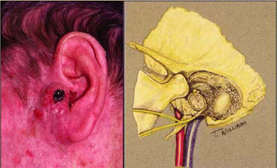
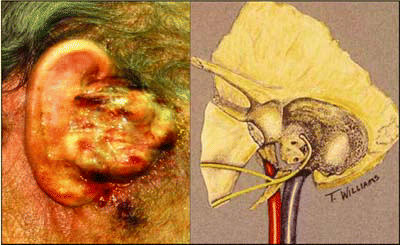
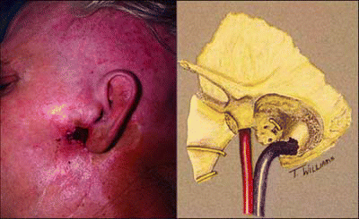
Tumors that can invade the temporal bone have three possible origins: the skin of the outer ear, the parotid gland, and the ear canal. Those that arise in the outer ear and parotid gland are the most common, whereas those arising in the ear canal are rare. Skin tumors present as a mass or ulcer in the outer ear, while parotid tumors present as a mass in the parotid gland. Tumors of the ear canal may present with signs that mimic chronic otitis media, such as hearing loss, purulence, or a canal mass. Other symptoms that can signal the presence of a tumor that invades the temporal bone are bleeding from the ear and facial paralysis or weakness.
“Any tumor that invades the temporal bone can produce purulent drainage, hearing loss, or affect the facial nerve,” said Paul W. Gidley, MD, Associate Professor at the Head and Neck Center at M. D. Anderson Cancer Center in Houston.
Diagnosis is made by physical examination of the head and neck, including thorough examination of the ear, parotid gland, neck, and cranial nerves and an evaluation of hearing. Imaging can assess the extent of the primary tumor and its relationship to adjacent structures, including the dura, brain tissue, facial nerve, and carotid artery. Imaging of the chest and the temporal bones and neck can assess the presence of metastases.
Often malignant tumors are misdiagnosed as an infection and treated with antibiotic drops. If a patient does not respond to treatment and has a mass in the ear canal, a biopsy should be obtained for diagnosis, said Barry Hirsch, MD, Professor of Otolaryngology at the University of Pittsburgh Eye and Ear Institute.
Preoperative Evaluation
Along with the overall health of the patient, the hearing status of the contralateral ear should be assessed. Staging should be done preoperatively by exam and imaging to provide some idea of the depth and invasion of the tumor. Both CT and magnetic resonance imaging provide complimentary information to assess the location and degree of tumor involvement. A CT scan can help determine whether bony erosion has occurred in the ear canal, whether the tumor has invaded the middle ear space, and if the tumor has spread to the temporomandibular joint. MRI can assess whether the parotid, upper neck lymph nodes, facial nerve, dura and brain, and soft tissues of the skull are involved, Dr. Hirsch explained.
Treatment
The terminology of surgery can be confusing, Dr. Hirsch continued. A sleeve resection refers to a procedure that removes the skin of the ear canal. For tumors limited to the external auditory canal, a lateral temporal bone resection is commonly performed. Following a complete mastoidectomy that is extended well into the zygomatic root, an en bloc resection of the bony external auditory canal and skin is done. This also entails removal of the tympanic membrane, malleus, and incus.
Treatment of tumors that invade the temporal bone is relatively straightforward but at the same time complex, Dr. Gidley continued. Tumors of the skin of the outer ear are typically squamous cell or advanced basal cell. These are treated with wide local excision, including temporal bone resection with parotidectomy and possibly lymph node dissection in the neck. Parotid tumors are also treated with lateral temporal bone resection and usually a total parotidectomy, attempting to preserve the facial nerve. Dr. Gidley said that parotid tumors often involve the facial nerve and a nerve graft may be necessary.
Also, these tumors are treated with selective neck dissection of the lymph nodes, depending on the pathology of the tumor and extent of invasion.
“Usually surgery for parotid tumors leaves a deficit in the facial nerve,” Dr. Gidley said.
Treatment of primary temporal bone tumors in the ear canal becomes more controversial as the tumors get larger, Dr. Gidley commented. Those that are confined to the ear canal are treated with lateral temporal bone resection, parotidectomy, and selective neck dissection. Tumors that invade the middle ear and mastoid bone are treated with a subtotal temporal bone resection combined with parotidectomy and selective neck dissection.
A total temporal bone resection involves removal of the carotid artery and is reserved for stage IV tumors, Dr. Hirsch said. A subtotal lateral temporal bone resection spares the carotid artery.
“If the tumor has eroded through the dura into the brain, surgery is contraindicated. Surgery under these conditions is not curative. Patients with severe pain should be managed with pain medications, chemotherapy, and radiotherapy,” Dr. Hirsch commented.
Dr. Hirsch said that removal of lymph nodes in the neck is controversial, because it is unclear whether a neck dissection improves survival. “In a patient with no clinical evidence of node involvement—that is, you can’t feel them and there is no suspicion on imaging—it is controversial whether to take the nodes out from the parotid gland and neck. If tumor is present, it will help determine the field of radiotherapy. Many surgeons perform a superficial parotidectomy, leaving it attached to the resected ear canal, which removes the first region of lymph node mestastases,” Dr. Hirsch said.
Very large tumors, whether originating in the skin, parotid gland, or ear canal, might be considered for preoperative chemotherapy. They are also frequently treated with postoperative radiation, Dr. Gidley said.
Postoperative radiation is also used when the primary tumor has surgical margins less than 5 mm or if the tumor is too close to the carotid artery or facial nerve to allow wide surgical margins, when the margins are microscopically positive, and if there is perineural invasion. Radiation is also used in most cases of adenoid cystic carcinoma and when there are multiple positive nodes or extracapsular extension of tumor.
Effects of Surgery
Postoperative radiotherapy can cause dry mouth. Surgery may cause paralysis of the facial nerve. Lateral temporal bone resection involves removal of the bony ear canal, tympanic membrane, and the small bones inside the ear, which destroys sound conductivity but preserves inner ear hearing, Dr. Gidley explained.
“Patients treated with lateral temporal bone resection have maximum conductive hearing loss. We can measure hearing in different ways to find the site of hearing loss. Older people usually have some degree of inner ear hearing loss, while those who undergo this type of surgery will have impaired conduction of sound,” he said.
Postoperative care includes hospitalization (possibly for up to two weeks), perioperative antibiotics, suction drains until output is less than 30 to 50 mL/day, and careful observation of the surgical wound for evidence of bleeding, cerebrospinal fluid leak, or flap necrosis.
Follow-up care depends on the extent of the surgery and the risk of recurrence. Appointments with the surgeon, radiation oncologist, and dentist are made on an individualized basis. In general, patients are seen anywhere from every month to every three months during the first year postsurgery, with longer intervals between appointments with each progressive year. By the fourth and fifth year after surgery, patients are usually seen every four or six months, and after five years they can be seen annually. All patients with malignant tumors should have annual chest X-rays and liver enzyme tests. Thyroid function tests are needed for patients whose lower neck received radiation.
Prognosis
Patients with stage IV tumors arising from the skin and parotid gland that invade the temporal bone tend to have a worse prognosis than those with smaller tumors arising from these sites. The average five-year survival rate is about 40%, Dr. Gidley said. Primary tumors of the temporal bone that are confined to the ear canal have a five-year survival of 80% to 100%, but if the tumor invades the mastoid bone or the inner ear, five-year survival drops to between 30% and 50%.
How Well Are Surgeons Doing?
A study presented at the recent meeting of the Triological Society showed that lateral temporal bone resections were effective for malignant tumors of the parotid gland, periauricular area, and the skin. The study was presented by Hung H. Dang, MD, a resident at the University of Oklahoma Health Sciences Center in Oklahoma City, OK.
“This type of surgery provides reasonable locoregional control and survival, and can be done with minimal morbidity to the patient,” stated Jesus Medina, MD, Professor and Chairman of the Department of Otolaryngology at the University of Oklahoma in Oklahoma City, and senior author of the study. Dr. Medina emphasized that good results are dependent on close collaboration between head and neck oncologic surgeons and otologists.
The retrospective study focused on 52 patients with malignant tumors adjacent to or invading the temporal bone who were treated with lateral temporal bone resection. The study included 43 males and nine females ranging from 30 to 91 years of age. Fifty percent of tumors were squamous cell, 25% were basal cell, and 23.1% were parotid carcinomas. There was one melanoma and one metastatic nasopharyngeal carcinoma.
In an interview, Dr. Medina said that these patients were treated with either subtotal resection to remove all of the temporal bone up to the internal carotid artery, or one of four variations of lateral temporal bone resections (Type I–IV). He noted that “total” is a misnomer, because not all of the temporal bone is removed with these procedures.
“We have been doing these procedures for quite some time at our institution. More often than not, tumors are attached to the temporal bone and may invade the mastoid. We found that we didn’t need to remove most of the bone, but could just remove areas of the bone adjacent to or actually invaded by tumor. The extent of surgery depends on involvement of the tumor,” Dr. Medina said.
Dr. Medina noted that results of the study should be viewed in the context of the patient population, which included mainly patients with advanced tumors. Sixty-nine percent of patients had previous treatment, including radiotherapy, and 28% already had facial paralysis when they underwent lateral temporal bone resection.
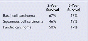
Local control was possible in 100% of 13 patients with basal cell carcinoma. Overall disease-free survival at two years was 46% for those with squamous cell carcinoma, 67% for basal carcinoma, and 50% for parotid carcinoma. At the last follow-up, 29 patients were alive and disease-free.
“We were able to preserve the inner ear in all patients. We were able to preserve the facial nerve in the majority of patients who did not have facial paralysis at baseline,” he continued.
Dr. Medina stated that the indications for lateral temporal bone resections and subtotal resection are different. The following types of tumors should have lateral temporal bone resection: tumors of the perauricular skin that extend to the auditory canal and the parotid gland immediately adjacent to, adhering to, or superficially invading the temporal bone but not invading the middle ear space or the aerated spaces of the mastoid. Indications for subtotal resection are when the tumor has invaded the middle ear space.
“For example, if a tumor of the pinna or parotid gland is up against the temporal bone or superficially invading the bone, lateral temporal bone resection should be performed. If the tumor invades the middle ear space or aerated spaces of the mastoid and if it does not surround the carotid artery, patients are eligible for subtotal resection,” he stated.
©2007 The Triological Society


Leave a Reply