- TLM Safe Salvage Option for T1a and T1b Glottic Cancers
- Post-Operative Pain and Bleeding Risk Following Tonsillectomy
- Tissue-Engineered Regeneration of Mastoid Air Cells Improves Eustachian Tube Function
- Two-Stage Process Repairs Internal Lining in Nasal Deformity
- Impact of Treatment Modality and Radiation Technique in Cancer Patients
- Psychological Impact of Wait Time for Thyroid Surgery
TLM Safe Salvage Option for T1a and T1b Glottic Cancers
What is the role of transoral laser microsurgery (TLM) for salvage in radiation failures for selected glottic cancers?
Background: TLM has replaced open laryngeal surgery for managing early glottic cancer, while preserving organ and function. It is also easily repeated for local recurrences, with better functional outcomes and shorter hospitalizations. This study was conducted to address TLM in the management of recurrent glottic cancer after failure of definitive radiation.
Study design: Retrospective analysis of medical records of 18 patients with recurrent T1 and T2 of the glottis after receiving radiation therapy. Records between 2002 and 2007 were examined.
Setting: Academic medical center in South Korea.
Synopsis: Only patients with recurrent glottic cancers were included in this study. All had received primary radiation and were re-staged at the time of recurrence with CT scans, endoscopy and biopsies due to a tendency to understage these recurrences initially. Only recurrent T1 and T2 cancers with adequate exposure were selected for TLM. A vestibulectomy was performed, followed by a CO2 laser en bloc or piecemeal excision. Frozen section of the margins was performed, and no patients underwent a neck dissection. The three- and five-year local control rates after TLM were 65 percent and 40 percent, respectively. Local control rates were much lower for those with anterior commissure involvement. Six of the 18 patients underwent salvage laryngectomies. There was a 90 percent disease-specific five-year survival after either salvage surgery or TLM.
Bottom line: Although the sample size was small, this retrospective study demonstrates that TLM is a relatively safe salvage option for recurrent T1a and T1b glottic cancers. For T2 and anterior commissure cancers, the high local recurrence rate makes TLM a less feasible option.
Reference: Han YJ, Lee SH, Kim SW, et al. Transoral laser microsurgery of recurrent early glottis cancer after radiation therapy: clinical feasibility and limitations. Ann Otol Rhinol Laryngol. 2012;121:375-382.
—Reviewed by Natasha Mirza, MD
Post-Operative Pain and Bleeding Risk Following Tonsillectomy
What is the association of post-operative pain and risk of hemorrhage after tonsillectomy?
Background: Tonsillectomies are among the most common medical procedures performed in the U.S., and their most common serious complication is post-operative hemorrhage. A high proportion of patients also suffer various degrees of post-operative pain. This study sought to investigate the association of post-operative pain behavior with post-operative hemorrhage.
Study design: This questionnaire-based study was conducted retrospectively on 335 patients and included patients undergoing both tonsillectomies and adenotonsillectomies. The incidence of hemorrhage and post-operative pain were evaluated retrospectively. Pain was graded using a visual analog scale at five time points and with five grades of severity.
Setting: Academic medical center in Klagenfurt, Austria.
Synopsis: Post-operative pain severity and duration were clustered into five levels of pain types. Hemorrhage included any episode of bleeding. Pain type V (very high or increasing pain for two weeks after surgery) had a statistically significant risk of hemorrhage (41 percent versus 18 percent for pain type I). There was also a strong relationship between indications for surgery and post-operative pain, with patients with recurrent tonsillitis and adult patients having higher pain levels. Children tended to have pain type I, whereas adults had pain types III-V.
Bottom line: This is the first study that has described an important association between post-operative tonsillectomy pain and bleeding. It is a well-conducted study that has emphasized the importance of paying close attention to the patient who complains of increasing pain one week following surgery.
Reference: Sarny S, Habermann W, Ossimitz G, Stammberger H. Significant post-tonsillectomy pain is associated with increased risk of hemorrhage. Ann Otol Rhinol Laryngol. 2012;121:776-781.
—Reviewed by Natasha Mirza, MD
Tissue-Engineered Regeneration of Mastoid Air Cells Improves Eustachian Tube Function
Can regenerated mastoid air cells (MACs) restore normal gas exchange function and contribute to improved Eustachian tube (ET) function?
Background: Most cases of chronic otitis media (OMC) are associated with poor development of the MACs and poor ET function. A common treatment for OMC is tympanoplasty with mastoidectomy; however, this procudure does not aim directly for the recovery of MACs and ET function. Further, recurrence of OMC after the procedure is common.
Study design: Clinical trial with control.
Setting: Department of Otolaryngology, The Foundation for Biomedical Research and Innovation, Kobe; Department of Otolaryngology-Head and Neck Surgery, Medical Research Institute, Kitano Hospital, Osaka; Department of Otolaryngology, Shizuoka General Hospital, Shizuoka; Department of Otolaryngology-Head and Neck Surgery, Graduate School of Medicine, Kyoto University, Kyoto; Department of Bioartificial Organs, Institute for Frontier Medical Sciences, Kyoto University, Kyoto, Japan.
Synopsis: Seventy-six patients with cholesteatoma, adhesive otitis media or OMC received tympanoplasty with mastoidectomy and MAC regeneration in a two-stage operation. During the first stage, artificial pneumatic bones and/or autologous bone fragments were implanted into the opened mastoid cavity. During the second stage, a nitrous oxide (N2O) gas study was performed in five patients with good MAC regeneration and five patients with poor MAC regeneration to measure middle ear pressure. For the control group, middle ear pressure was measured in five patients with well-developed MACs during cochlear implantation or facial nerve decompression. ET function was also measured twice in each patient, before the first operation and six months after the second.
At the second-stage operation, in all cases with regenerated MACs and in the normal control group, middle ear pressure changed after administration of N20. In contrast, no change was observed in cases with unregenerated MACs. In 70 percent of the regenerated MAC group, ET function was improved, whereas improvement of ET function was observed in only 13 percent of the unregenerated MAC group.
Bottom line: Tissue-engineered regeneration of MACs improves ET function and gas exchange in the middle ear.
Reference: Kanemaru S, Umeda H, Yamashita M, et al. Improvement of Eustachian tube function by tissue-engineered regeneration of mastoid air cells. Laryngoscope. 2013;123:472-476.
—Reviewed by Sue Pondrom

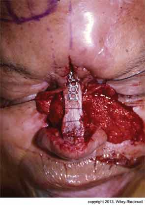
Two-Stage Process Repairs Internal Lining in Nasal Deformity
When septal hinge flaps are unavailable, what method can be used to reconstruct large defects of internal lining in a full-thickness nasal deformity?
Background: Full-thickness nasal deformities are a reconstructive challenge and require reconstitution of external skin, internal lining and structural support. Restoration of a reliable internal lining is critical, with septal hinge flaps often used. However, sometimes these and other intranasal mucosal flaps are unavailable.
Study design: Case study.
Setting: Department of Otolaryngology-Head and Neck Surgery, University of Michigan Health System, Ann Arbor.
Synopsis: Surgeons described the use of a two-stage interpolated subcutaneous fat pedicle melolabial flap to reconstruct large defects of internal lining when septal hinge flaps were unavailable. They noted that this technique is particularly useful to salvage patients with full-thickness nasal defects who have already expended their first forehead flap. The patient was a 69-year-old male who had undergone radiation and nasal reconstruction where the pericranial flap failed and the split calvarial bone became exposed and was lost, creating a large fistula. The authors of this study performed a second reconstructive surgery using a 5 x 3-cm two-staged subcutaneous fat pedicle melolabial flap. They noted that the potential drawbacks of the interpolated melolabial flap are that it lacks a true axial vascular pedicle and that it may be hair-bearing in men. However, their patient had no problems with excessive hair growth and, in the four years since this second operation, he has continued to do well.
Bottom line: A two-stage interpolated subcutaneous fat pedicle melolabial flap reconstructed a near-total nasal lining defect in a patient with unavailable septal hinge flaps, offering an alternative to using a forehead flap, and may be an alternative to a free radial forearm flap in select patients.
Reference: Griffin GR, Chepeha DC, Moyer JS. Interpolated subcutaneous fat pedicle melolabial flap for large nasal lining defects. Laryngoscope. 2013;123:356-359.
—Reviewed by Sue Pondrom
Impact of Treatment Modality and Radiation Technique in Cancer Patients
What is the impact of chemoradiation (CRT) and radiation techniques on toxicity, quality of life (QoL) and functional outcome in hypopharyngeal cancer patients?
Background: In addition to curing the patient, the goals for treatment of hypopharyngeal cancer (HPC) should include laryngeal preservation while minimizing side effects. Patients with T3 and early T4 can be offered the possibility of organ preservation with CRT, but there are high rates of treatment-related toxicity. Inconsistent reports exist regarding treatment-related toxicities and assessment of QoL for this treatment.
Study design: Retrospective analysis.
Setting: Department of Radiation Oncology, Department of Biostatistics, and the Department of Otorhinolaryngology, Erasmus MC-Daniel den Hoed Cancer Center, Rotterdam, Netherlands.
Synopsis: The authors looked at 176 consecutive patients with HPC at one institution from 1996 to 2010. All were treated with CRT or radiotherapy (RT) alone. End points were acute and late toxicity, QoL, assessment and functional outcome using laryngoesophageal dysfunction-free survival (LED-FS), defined by the Larynx Preservation Consensus Panel. All patients experienced one or more acute side effects, with the most serious being grade 3 skin toxicity, mucosal toxicity and dysphagia. Fourteen percent of patients needed hospitalization for severe mucositis, dysphagia and weight loss; intercurrent infection; or severe malaise.
CRT, compared with RT alone, increased the overall rate of grade 3 acute toxicity; however, significantly less grade 3 acute dermatitis, mucositis and pain were observed in patients treated with intensity-modulated radio therapy (IMRT) compared with three-dimensional conformal radiotherapy (3DCRT). Fifty-seven patients experienced one or more types of late toxicity grade 2 or higher, with dysphagia and xerostomia the most common. Use of IMRT, compared with 3DCRT, significantly reduced incidences of late complications. Eighty-three percent of patients with grade 3 acute toxicity later developed grade 3 late toxicity, and 40 percent of patients were feeding-tube dependent at the end of treatment. Regarding functional outcome, 44 percent of patients were still alive at the last follow-up. Of these, 65 patients had organ preservation. All of the patients eligible for QoL analysis had been treated by IMRT, so QoL scores could not be evaluated; however, the deterioration in scores was more pronounced in patients treated by CRT compared to RT alone. QoL scores on all scales deteriorated during treatment.
Bottom line: When compared with radiation alone, CRT significantly improved LED-FS as a suitable indicator for functional outcome. CRT was noted to increase acute, but not late, radiation-induced side effects without deterioration in QoL scores. Patient-reported xerostomia was more pronounced in patients treated by CRT, but this was not translated as significant deterioration on the functional scales. When compared with 3DCRT, IMRT significantly reduced acute and late toxicity.
Reference: Al-Mamgani A, Mehilal R, van Rooij PH, Tans L, Sewnaik A, Levendag PC. Toxicity, quality of life, and functional outcomes of 176 hypopharyngeal cancer patients treated by (chemo)radiation: the impact of treatment modality and radiation technique. Laryngoscope. 2013;122:1789-1795.
—Reviewed by Sue Pondrom
Psychological Impact of Wait Time for Thyroid Surgery
What is the degree of psychological morbidity in patients waiting for thyroid surgery?
Background: Waiting for health care is an important source of anxiety for Canadians. As of 2005, the province of Ontario has implemented a wait time system to improve access to surgical services: priority level 1 for immediate/urgent; priority level 2 for fewer than 14 days; priority level 3 for fewer than 28 days; and priority level 4 for fewer than 84 days. Most patients with thyroid cancer or indeterminate thyroid nodules are categorized as priority level 4. The degree of psychological distress patients experience during this wait is unknown.
Study design: Prospective assessment of patients’ pre- and post-operative psychological morbidity.
Setting: Department of Otolaryngology-Head and Neck Surgery, Department of Psychosocial Oncology and Palliative Care, Department of Endocrinology, University Health Network, Ontario Cancer Institute, University of Toronto; Department of Otolaryngology-Head and Neck Surgery, Mount Sinai Hospital; Department of General Surgery, Toronto General Hospital; Department of Surgical Oncology, Department of Otolaryngology-Head and Neck Surgery, Princess Margaret Hospital; Department of Otolaryngology-Head and Neck Surgery, Sunnybrook Health Sciences Center, Toronto, Ontario.
Synopsis: Patients waiting for thyroidectomy were mailed a sociodemographic and four psychological morbidity questionnaires: Impact of Event Scale-Revised (IES-R), Illness Intrusiveness Ratings Scale (IIRS), Perceived Stress Scale (PSS) and Hospital Anxiety and Depression Scale (HADS). Over a three-year period, the response rate was 53 percent for 176 patients providing pre-operative data and 42 percent completing post-operative data. Respondents with a suspicious or known malignancy waited an average of 107 days for thyroidectomy, while those with benign neoplastic biopsies waited an average of 218 days. Although respondents reported substantial psychological morbidity on all questionnaires, there was no significant association between psychological morbidity and wait times, clinical or sociodemographic factors. Post-operative anxiety decreased significantly for all measures except for the IIRS.
Bottom line: Patients waiting for thyroid surgery have mild to moderate psychological morbidity and long wait times for surgery, but these appear to be unrelated. The anxiety decreases after surgery.
Reference: Eskander A, Devins GM, Freeman J, et al. Waiting for thyroid surgery: a study of psychological morbidity and determinants of health associated with long wait times for thyroid surgery. Laryngoscope. 2013;123:541-547.
—Reviewed by Sue Pondrom
Leave a Reply