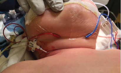
Fig. 1. Electrode needles of additional active leads are placed into the neck close to the thyroid cartilage.
Explore This Issue
April 2021© Lawlor, et al. Laryngoscope.
Introduction
The recurrent laryngeal nerve (RLN) is at risk during pediatric surgery of the neck, mediastinum, and chest. RLN injury during surgery for esophageal atresia, tracheoesophageal fistula, and tracheobronchomalacia (TBM) is established in the literature as the RLN often courses through the operative field. Identifying and protecting the RLN is particularly difficult in the surgery of neonates, aberrant anatomy, and in reoperative cases (J Pediatr Surg. 2019;54:1551-1556; Laryngoscope. 2020;130:E65–E74).
Intraoperative nerve monitoring (IONM) is standard of practice in adult thyroid surgery but has not yet been widely adopted in pediatric surgery (Laryngoscope. 2011;121:S1-S16). An obstacle to routine utilization of IONM in pediatric surgery is the lack of size-appropriate nerve monitoring devices for the pediatric patient. Translaryngeal and endolaryngeal electrodes may be used in small infants, but they are associated with significant challenges. Translaryngeal electrodes may obstruct the surgical field. Endolaryngeal electrodes may be difficult to place, cause trauma, or dislodge with positioning or intraoperative bronchoscopy. Endotracheal tubes (ETTs) with integrated or adhesive electrodes are the most common way to monitor the RLN. Single channel adhesive electrodes are available for ETT as small as 2.0 mm inner diameter (ID) but do not allow for monitoring of each nerve individually. Thus, if one nerve is injured intraoperatively, the surgeon may not be alerted by the system because the contralateral nerve is functioning. Double-channel electrodes allow for monitoring of each nerve individually. Unfortunately, integrated and adhesive double-channel electrodes (to monitor each RLN separately) are only available in ETT appropriate for children aged 4 years or older. We present a modification of a double-channel adhesive electrode for IONM of the RLN in children as young as term infants.
Method
Surgical Technique. For all cases of intraoperative RLN monitoring, chemical paralysis must be avoided and preoperative communication between the surgical and anesthesia teams is vital. An appropriately sized standard ETT is selected for the patient down to a size 3.0 mm ID (term infant). As the electrode slightly increases the outer diameter of the ETT, the ETT may need to be downsized to prevent excessive pressure on the larynx and subglottis. A Neurovision Dragonfly Stick-On double-channel electrode of small size is then custom trimmed to fit the smaller ETT. The Dragonfly adhesive electrodes are compatible with both Neurovision’s Nerveana Nerve Locator and Monitor system and the Medtonic Nerve Integrity Monitoring (NIM) systems.
The Dragonfly double-channel sensor is trimmed by the surgeon, otolaryngologist, or anesthesiologist. Depending on the size of the ETT, there are two different ways to trim the sensor. For a smaller ETT (3.0 or 3.5), the two lateral electrodes are trimmed such that the two center channels or electrodes remain in line with the adhesive tape, resulting in a significantly narrower double-channel electrode. For an ETT sized 4.0 or 4.5, a single lateral electrode is cut off, leaving three active electrodes on the sensor (two of the same color). Regardless of how the sensor is trimmed, a voltmeter is then used to determine which leads are active. The inactive leads are marked by placing a small knot on them to avoid connecting them to the NIM system. If a voltmeter is not available, one can unravel the cords to determine which electrodes are active. Once the active leads are determined, the trimmed sensor is wrapped around the desired ETT. Care is taken to place the electrode proximal to the cuff (if using a cuffed tube) at the approximate level that the vocal folds will lie after intubation.
An obstacle to routine utilization of intraoperative nerve monitoring in pediatric surgery is the lack of size-appropriate nerve monitoring devices for the pediatric patient.
The patient is then intubated by the anesthesia or surgical team using a concurrent media access control blade. The two horizontal blue lines on the sensor help to mark the midpoint of the electrodes which should contact the true vocal folds. The tube is then secured by anesthesia. The electrode needles of the additional (one or two depending on how the sensor was trimmed) active electrodes (to replace those tied off and not used on the Dragonfly) are placed into the anterior neck near the thyroid cartilage to ensure function of the double-channel system (Fig. 1). Two grounding leads (one for the surface electrode system and one for the stimulator) are placed into muscle such as the deltoid or trapezius. The Dragonfly electrode is then connected to the nerve monitoring system of choice and a baseline amplitude should read for each vocal fold individually. Function of the system is ultimately confirmed by stimulation of each nerve individually with a nerve probe once the surgical field is open. It is important to note that the nerve monitoring systems will still function after the electrode has been modified if it is set up appropriately, though this is not a manufacturer-approved modification.
Troubleshooting. One must recognize that there is a substantial learning curve to the successful implementation of this technology (World J Surg. 2020 Jun;44:1874-1875). Patience and persistence are often needed to troubleshoot the system. One must be able to understand all the components of the nerve monitoring system in order to troubleshoot them in a systematic way. We propose the following:
- No signal: Check all electrodes are connected appropriately and that impedance levels are less than 1 Ω. If impedance levels are greater than one then that particular electrode needs to be placed closer to the operative field and/or deeper within the tissues. Perform video-laryngoscopy to ensure sensors are truly at the level of the vocal cords. Make sure receiver box has a working fuse.
- Nerve will not stimulate: Perform a video laryngoscopy to ensure the electrodes are positioned on the vocal folds.
- Excessive background noise: Ensure silencer is wrapped around electrocautery cord.
- Loss of signal: Evaluate entire system.
Results
A 4-month-old girl with severe TBM and brief resolved unexplained events underwent posterior tracheopexy, bilateral bronchopexies, rotation esophagoplasty, and partial thymectomy via right thoracotomy. Bilateral RLNs were at risk during the procedure. A Dragonfly stick-on double-channel electrode was modified as described above and wrapped around a 3.5 cuffed ETT. Function of the nerve monitoring system was confirmed with stimulation of bilateral RLN using a nerve probe intraoperatively. Her preoperative and postoperative flexible fiberoptic laryngoscopy showed normal vocal fold movements bilaterally.