INTRODUCTION
Chondrolaryngoplasty is a procedure designed to reduce a conspicuous laryngeal prominence and feminize the neck. Traditionally, this procedure is performed through a transcervical incision, which may lead to an unsightly visible scar that can “out” the patient. To address this, indirect approaches have been employed utilizing a submental incision, which can be concealed by the chin. This approach nevertheless has the result of a visible neck scar and carries the risk of tethering to the underlying cartilage framework. To address this issue, Khafif et al. described scarless chondrolaryngoplasty through a transoral endoscopic vestibular approach (TOEVA) (Facial Plastic Surg Aesthet Med. 2020;22:172–180).
One of the key aspects of performing a safe chondrolaryngoplasty is the ability to identify the location of the anterior commissure tendon. Destabilizing the anterior commissure tendon by overaggressive resection can significantly and irreversibly affect the patient’s voice (Plast Reconstr Surg Glob Open. 2018;6:e1877). We describe a modification of the approach described by Khafif et al. in which we use a suture to mark the anterior commissure. We modified the technique described by Spiegel et al. in which a 22-gauge needle is used to mark a location 2 mm superior to the insertion of the anterior commissure tendon before performing the cartilage resection (Arch Otolaryngol Head Neck Surg. 2008;134:704–708). In our technique, we use a 22-gauge spinal needle and thread a 3-0 prolene suture through the needle lumen while directly visualizing the larynx. This allows precise localization of the inferior limit of resection of cartilage continuously throughout the procedure. Institutional review board sanction was obtained, and this case report was considered exempt from formal approval.
METHOD
A 21-year-old trans woman with no past medical history presented to our clinic with gender dysphoria. She had a history of facial feminization surgery including rhinoplasty and sliding genioplasty but remained dissatisfied with the appearance of her neck. Physical examination revealed a protuberant laryngeal prominence.
The patient was offered a chondrolaryngoplasty. We discussed the traditional transcervical approach, the submental incision approach, and the TOEVA. Given concerns over cosmetic appearance of a neck scar, the patient strongly preferred the scarless approach. The patient was intubated with a reinforced size 6-0 endotracheal tube. Unasyn was administered as intraoperative prophylaxis. A point midway between the thyroid notch and the inferior border of the thyroid cartilage was demarcated. A direct laryngoscopy was performed,and the patient was placed into suspension. The anterior commissure was visualized, and externally a 22-gauge spinal needle was used to puncture through the previously demarcated central portion of the thyroid cartilage and through the anterior commissure in the endolarynx. A size 3-0 prolene was passed through the needle lumen and grasped in the endolarynx. The laryngoscope was removed and the suture ends were secured externally using clamps.

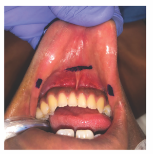
Figure 1. The vestibular incisions are plotted. The central gingivobuccal incision is 1 cm anterior to the labial frenulum of the lower lip and spans 1.5 cm. Two stab incisions are plotted near bilateral oral commissures.
Three incisions were marked and infiltrated with 1% lidocaine in 1:100,000 epinephrine: a central gingivobuccal incision 1 cm anterior to the labial frenulum of the lower lip measuring 1.5 cm, and two stab incisions near the oral commissures (Figure 1). Mucosal incisions were made using a 15-mm blade, and Crile and Kelly clamps were used to develop the subplatysmal flap plane in the midline incision. Hegar dilators were used in the central pocket to dilate the subplatysmal tract to 5 mm. A 5-mm trochar was then placed in the central incision and two 5-mm ports were placed in the lateral incisions. The subplatysmal plane was developed under direct visualization with a 0-degree scope using a harmonic device, and the pocket was insufflated with CO2 to 6 mmHg. The prolene suture was noted and carefully avoided.
A 30-degree scope was used, and the strap muscles were separated superior to the region of the prolene suture. Dissection was performed to identify the thyroid notch and thyrohyoid membrane. Inferiorly, dissection ended at the level of the prolene suture, which served to demarcate the level of the anterior commissure of the true vocal cords. A laparoscopic L-hook bovie was used to cauterize the superior aspect of the thyroid cartilage, releasing the perichondrium. The perichondrium of the anterior aspect of the laryngeal prominence was dissected bluntly with the laparoscopic Endo Peanut dissector to the level of the prolene suture, and the posterior perichondrium was dissected using a laparoscopic spatula elevator to the level of the thyro- epiglottic ligament. Curved endo-scissors were used to remove the prominent laryngeal prominence, with the incision starting just anterior to the oblique line of the thyroid cartilage and carried anteriorly to a point 3 mm superior to the prolene suture. The Sonopet ultrasonic aspirator at a power of 100%, suction of 50%, and irrigation of 15 ml/min was used to sculpt protruding edges of cartilage. The L-hook bovie was used to reduce bunched perichondrium in the inferior border of the excision. The prolene was removed and wound bed was irrigated. A barbed self-locking 4-0 monocryl suture was used to close the strap musculature. Five milliliters of Evicel was placed under direct visualization. The trocars were removed, and the oral incisions were closed with running locking sutures using 4-0 chromic. A final postoperative photograph was performed, demonstrating decreased projection of the laryngeal prominence. Fluffs and Tensoplast were placed over the cervicomental angle as a pressure dressing.
RESULTS
Postoperatively, the patient was sent home after routine observation. The pressure dressing was removed after 24 hours. Discharge medications included standing Tylenol, amoxicillin clavulanate, and a short course of opioid medications. The patient was instructed to follow a soft diet until her first postoperative visit. At her postoperative visit, her intraoral incisions were healing well and she was without pain or any complications.
We have herein described a modification to this approach with a novel technique to mark the anterior commissure tendon. This marking suture is clearly visible throughout the endoscopic portion of the procedure, allowing delineation of the inferior border of resection of cartilage. The total anesthesia time was 4 hours and 34 minutes, and total surgical time was three hours and 24 minutes. Postoperatively, our patient was very pleased with her neck cosmesis.