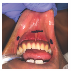
Figure 1. The vestibular incisions are plotted. The central gingivobuccal incision is 1 cm anterior to the labial frenulum of the lower lip and spans 1.5 cm. Two stab incisions are plotted near bilateral oral commissures.
Three incisions were marked and infiltrated with 1% lidocaine in 1:100,000 epinephrine: a central gingivobuccal incision 1 cm anterior to the labial frenulum of the lower lip measuring 1.5 cm, and two stab incisions near the oral commissures (Figure 1). Mucosal incisions were made using a 15-mm blade, and Crile and Kelly clamps were used to develop the subplatysmal flap plane in the midline incision. Hegar dilators were used in the central pocket to dilate the subplatysmal tract to 5 mm. A 5-mm trochar was then placed in the central incision and two 5-mm ports were placed in the lateral incisions. The subplatysmal plane was developed under direct visualization with a 0-degree scope using a harmonic device, and the pocket was insufflated with CO2 to 6 mmHg. The prolene suture was noted and carefully avoided.
Explore This Issue
December 2022A 30-degree scope was used, and the strap muscles were separated superior to the region of the prolene suture. Dissection was performed to identify the thyroid notch and thyrohyoid membrane. Inferiorly, dissection ended at the level of the prolene suture, which served to demarcate the level of the anterior commissure of the true vocal cords. A laparoscopic L-hook bovie was used to cauterize the superior aspect of the thyroid cartilage, releasing the perichondrium. The perichondrium of the anterior aspect of the laryngeal prominence was dissected bluntly with the laparoscopic Endo Peanut dissector to the level of the prolene suture, and the posterior perichondrium was dissected using a laparoscopic spatula elevator to the level of the thyro- epiglottic ligament. Curved endo-scissors were used to remove the prominent laryngeal prominence, with the incision starting just anterior to the oblique line of the thyroid cartilage and carried anteriorly to a point 3 mm superior to the prolene suture. The Sonopet ultrasonic aspirator at a power of 100%, suction of 50%, and irrigation of 15 ml/min was used to sculpt protruding edges of cartilage. The L-hook bovie was used to reduce bunched perichondrium in the inferior border of the excision. The prolene was removed and wound bed was irrigated. A barbed self-locking 4-0 monocryl suture was used to close the strap musculature. Five milliliters of Evicel was placed under direct visualization. The trocars were removed, and the oral incisions were closed with running locking sutures using 4-0 chromic. A final postoperative photograph was performed, demonstrating decreased projection of the laryngeal prominence. Fluffs and Tensoplast were placed over the cervicomental angle as a pressure dressing.