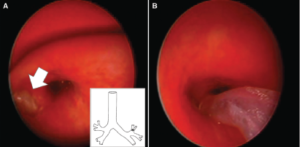
Figure 1. Bronchoscopic view during foreign body removal. (A) Peanut fragment seen lodged into the superior division of the left superior lobar bronchus (Inset *). (B) A 6-Fr flexible suction catheter with the tip bent was used to access the foreign body.
Figure 1. Bronchoscopic view during foreign body removal. (A) Peanut fragment seen lodged into the superior division of the left superior lobar bronchus (Inset *). (B) A 6-Fr flexible suction catheter with the tip bent was used to access the foreign body.
ENTtoday - https://www.enttoday.org/article/how-to-catheter-guided-basket-removal-of-a-difficult-to-reach-pediatric-airway-foreign-body/ent_1221_pg11a/
