INTRODUCTION
Frontal sinus (FS) surgery still represents a challenge due to its complex and highly variable anatomy. Several tumoral and infective pathologies, as well as traumatic injury, may affect this region, with possible intracranial and orbital involvement. Osteoma is the most common benign tumor that affects this area, followed by the inverted papilloma (Laryngoscope. 2012. doi:10.1002/lary.23275). The malignancy of FS is led by squamous cell carcinoma, representing 40%–60% of the sinonasal malignancies, followed by adenocarcinoma (Laryngoscope. 2015. doi:10.1002/lary.25465).
Explore This Issue
December 2023The endoscopic frontal sinus procedures present several limitations associated with individual anatomy and tumor features, especially in cases of lesions located superior and laterally within the frontal sinus (Ann Otol Rhinol Laryngol. 2012. doi:10.1177/000348941212100802).
Before the endoscopic era, the standard FS surgery involved a bicoronal incision. This technique required a large scalp flap, however, and could result in potential facial palsy, poor cosmetic results, and prolonged recovery.
The eyebrow approach is commonly used because it provides direct access to the frontal sinus with avoidance of an obvious facial scar. Besides the risk of eyebrow alopecia, one limitation is the supraorbital nerve injury, which normally limits the extension of the incision and the access to the frontal sinus. The eyebrow incision is traditionally performed medially or laterally to the supraorbital foramen to avoid injury to the supraorbital nerve (Arch Craniofac Surg. 2016. doi:10.7181/acfs.2016.17.4.186.).
To the best of our knowledge, there is no description in the literature of a surgical technique that utilizes the full extension of the eyebrow with preservation of the supraorbital nerve. In this study, we present a detailed anatomical description of an eyebrow approach that allows full exposure of the frontal sinus with a large osteoplastic bone flap and preservation of the supraorbital nerve. A clinical case is described to illustrate the surgical technique.
METHOD
Three embalmed and latex-injected cadaveric heads were dissected to assess the feasibility and limitations of the approach in documenting key steps. After the dissection, the technique was performed in a patient with an inverted papilloma of the frontal sinus.
Surgical Technique
After palpation of the supraorbital foramen, a skin incision was performed laterally to the foramen in the superior aspect of the eyebrow and extended to the lateral edge of the brow. This incision was performed with a 15-blade oriented in a 45-degree angle to the skin parallel to the hair follicles, decreasing the risk of extensive eyebrow alopecia. After the identification of the orbicularis oculi, the incision was extended medially to find the supraorbital nerve arising from the supraorbital foramen. The neurovascular bundle was dissected and isolated. The nerves were carefully dissected from the surrounding tissue, allowing superior retraction of the incision. The anterior wall of the frontal sinus was exposed medially and laterally to the supraorbital nerve. A navigation system was used to mark the limits of the anterior table of the frontal sinus. An anterior table osteotomy was performed with a bone cutting tip of the Sonopet Omni Ultrasonic Surgical System along the limits of the left frontal sinus. A 1-mm drill bit can be used for the osteotomy as well. The inferior bone cut was placed just superior to the supraorbital rim and the nerve was protected with a retractor. Subsequently, the anterior table was gently elevated and removed to expose the frontal sinus.
Illustrative Case
A 77-year-old male with a history of recurrent left-sidedfrontoethmoidal inverted papilloma underwent a first endoscopic resection in August 2020 and a second endoscopic surgery in June 2022, both performed at another institution. MRI and CT scan showed the recurrent tumor filling the left frontal sinus. Therefore, an open approach through an eyebrow incision was indicated to complete surgical resection.

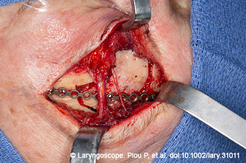
Figure 1. Intraoperative details of the approach and tumor excision. Pictured here is the bone flap reposition and fixation.
An osteoplastic bone flap of the anterior table of the frontal sinus was performed. There was tumor attached to the anterior table of the frontal sinus (bone flap), which was successfully removed. The inner surface of the bone flap was also drilled with a diamond burr to ensure complete tumor resection from the anterior table. The supraorbital nerve was preserved. Complete resection of the tumor was performed, with extirpation of the entire mucosa of the frontal sinus. A high-speed drill with a diamond burr was used to drill out the frontal sinus bony walls and to ensure complete tumor and mucosal extirpation.
During the cauterization of the posterior table of the FS, there was an area of small dural dehiscence and a cerebrospinal fluid (CSF) leak was observed. Dural synthetic substitute was placed through the opening and immediately stopped the CSF flow. A nasal floor free mucosal graft was also placed to reconstruct the defect. (The management of the intraoperative CSF leak is shown in the intraoperative video). Finally, the anterior table was repositioned and fixed with a titanium plate and microscrews.
No postoperative complications were reported, and the patient was discharged 24 hours after surgery with mild forehead hypoesthesia that recovered completely in three weeks.
RESULTS
The anatomical dissection showed the feasibility of the surgical technique. It allowed enough space for the osteotomy of the entire anterior table of the frontal with preservation of the supraorbital nerve. The lateral, superior, and medial limits of the frontal sinus, including the frontal sinus recess, were well exposed with this approach.
Some anatomical features and extension of the disease may limit the application of an endoscopic eyebrow approach. Frontal sinus anteroposterior dimension below 0.8 cm represents a limitation due to the acute angle and difficult surgical visualization. A pronounced convexity of the posterior wall of the frontal sinus makes it more difficult to manipulate the tumor, reducing intrasinus surgical maneuverability (Ann Otol Rhinol Laryngol. 2012. doi:10.1177/000348941212100802). ]
Moreover, the involvement of the posterior table of the FS entails the risk of CSF leak. As mentioned in the case description, CSF leakage was observed intraoperatively after the removal of the tumor that was successfully repaired through the open approach. Superior located frontal sinus CSF leaks are challenging for endoscopic skull base reconstruction.
In the three cadaveric specimens, the lateral limit of the FS extended lateral to the supraorbital nerve. The degree of exposure was the same as what is obtained with the bicoronal approach. The clinical case illustrated a successful application of this technique.