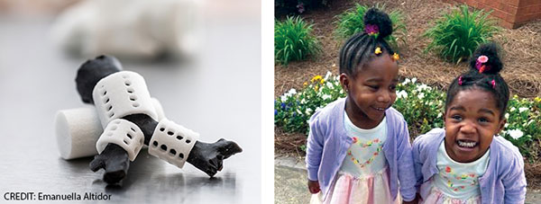
Justice, top, received 3D-printed tracheal splints (left), printed by the Georgia Institute of Technology, and now, she and her twin, Journee, are thriving.
Of course, tracheostomies and long-term ventilation aren’t ideal solutions. Tracheostomies require careful care and maintenance; positive pressure ventilation can cause “ongoing trauma to the lungs,” Dr. Maher said. Tracheostomy tubes and ventilators also inhibit children’s vocal and motor development.
Explore This Issue
October 2024
To the FDA’s credit, they recognize that getting a device like this fully approved may take 10 years, sometimes longer. So, if there is someone with critical health issues who could potentially benefit from the device, the FDA is willing to review each individual case.” — Kevin Maher, MD D
As a result, physicians sometimes use other approaches. During a tracheopexy, surgeons stabilize the floppy areas of the trachea by securing it to other nearby organs or tissues; these attachments prevent the trachea from collapsing as the affected individual breathes. It’s an effective, but far from ideal, treatment for severe tracheomalacia, as it requires operating around major blood vessels in a small chest cavity.
Some surgeons use intratracheal stents to hold the airway open. Intraluminal metal stents can be inserted endoscopically—a major advantage over open chest surgery. And evidence to date suggests that intraluminal stenting can be effective in children with tracheomalacia. In a 2023 paper describing the results of airway stenting with balloon-expandable coronary bare-metal stents to treat tracheobronchial disease in medically complex infants with congenital heart disease, six of eight treated patients were able to wean from mechanical ventilation within days of stent placement. The stents were removed approximately six months later, and bronchoscopy after stent removal “demonstrated a rounder configuration of the airway consistent with bronchial remodeling” (J Soc Cardiovasc Angiogr Interv. 2023;2:101068).
But Dr. Brigger, one of the authors of the paper, notes that internal airway stents “come with their own set of complications,” including the possible buildup of granulation tissue, which may occlude the airway and erosion of the stent through the walls of the airway.
External airway splints avoid those complications but require open chest surgery. The first report of external pediatric airway splinting using patient-specific 3D-printed splints was published in Science Translational Medicine in 2015. The study described positive results using bioabsorbable custom-printed airway splints to treat three children, ages three months, 16 months, and five months at the time of surgery. All three children had life-threatening tracheobronchomalacia and were confined to the hospital, dependent on mechanical ventilation via a tracheostomy prior to surgery. All three experienced significant improvement in their symptoms; one child no longer required ventilator support and was discharged home just three weeks after surgery (Sci Transl Med. 2015;7:285ra64).
A 2021 study reported on the results of implantation of external moldable, bioresorbable stents in 14 severely symptomatic children ages two months to 14 years. Seven of the children had tracheostomies in place at the time of surgery; six required mechanical ventilation in monitored, inpatient settings. Post-surgery, five patients were discharged home; two died of unrelated causes. Three children still required mechanical ventilation (JTCVS Tech. 2021;8:160-169).
When Justice continued to have breathing difficulties after surgical repair of her congenital aortic arch, Drs. Maher and Goudy and her parents discussed the possibility of supporting her airway via custom-printed 3D splints.
As otolaryngologists, we need to continue to tell the story of the unmet needs of our patients. We need to communicate that to engineers and scientists and work alongside them to develop new therapies.” — Stephen Goudy, MD