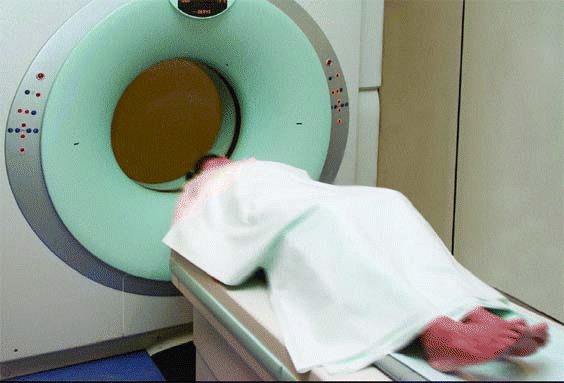
SAN DIEGO-With improved technology, as well as increased availability and access, diagnostic imaging has become the fastest growing segment of health care spending, with estimates of 15% to 35% increases annually. As a result, concerned third-party payers have established radiology guidelines, and some have teamed with radiology benefit management companies (RBMs) to bring down costs.

In the field of otolaryngology, the most common imaging service used is computed tomography (CT), considered by many to be the gold standard diagnostic study for imaging of the paranasal sinuses for inflammatory disease.
According to Medicare databases, there’s been an increase of 61 percent in the use of CT studies from 2001 to 2005, said Brent Senior, MD, Associate Professor of Otolaryngology-Head and Neck Surgery at the University of North Carolina, Chapel Hill. In the same time period, otolaryngology usage increased 195 percent.
Dr. Senior’s remarks came during an April 27 panel presentation on CT scanning of the paranasal sinuses held during the American Rhinologic Society’s (ARS) program at the Combined Otolaryngology Spring Meetings.
Referring to radiology cost management, Dr. Senior noted that while this process of imaging service management is, in and of itself, not inherently flawed, the primary end point should not be cost savings, but improved patient outcomes. Cross-specialty cooperation in the development of evidence-based criteria for imaging services is essential to ensure that patients have access to proper diagnosis.
The usage patterns for CT in otolaryngology were the topic of panelist Pete Batra, MD, Assistant Professor of Surgery in the Section of Nasal and Sinus Disorders at the Head and Neck Institute of the Cleveland Clinic Foundation. Discussing the results of a Web-based, 20-question ARS survey, he noted that there is a wide variation in how we scan patients when they come into our offices.
In general, the survey showed a general trend of increased CT usage from pretreatment to post-first and second rounds of treatment. The most commonly used for diagnosis were 3-mm coronal (with and without axials), with plain films rarely used. MRI was utilized by only 32% of responders.
Additionally, the majority of physicians surveyed said they utilized hospital radiology or free-standing imaging centers for CT, with few indicating they had an in-office CT scanner or financial interest in an imaging center. Forty-six percent who own an in-office CT scanner reported reading, interpreting, and billing for the scans.
Efficacy of Testing
The question then arises: Do all the tests ordered by otolaryngologists really add to improvement in patient diagnosis and treatment?
Rakesh K. (Rick) Chandra, MD, Clinical Assistant Professor at the University of Tennessee College of Medicine in Memphis, discussed currently accepted indications for CT scans, questionable circumstances for ordering imaging services, and research studies regarding CT scan effectiveness in diagnosis.
Most of us will probably agree that CT is not necessary for routine diagnosis and treatment of acute sinusitis, he said, because the CT doesn’t distinguish acute sinusitis from a viral upper respiratory infection. Most of us base our decision to treat with antibiotics, based on symptoms and the time course.
He added that physicians will obtain a CT scan frequently when there are any suspected orbital complications, if there is pending or suspected intracranial complication, if the diagnosis is in doubt, or if the patient fails consecutive courses of appropriate antibiotics. Additionally, a CT would probably be ordered for a suspected neoplasm, chronic rhinosinusitis of more than 12 weeks that has failed maximal medical therapy, polyposis, and suspected allergic fungal sinusitis (AFS) or mucocele.
The when-to-order dilemma, he said, occurs with patients under the following circumstances:
- Patients with suspected chronic rhinosinusitis who have negative CTs. He referred to a 2006 study where only dysosmia and polyps correlated with a positive CT scan.
- Patients without chronic rhinosinusitis often have positive CTs. A New England Journal of Medicine study in 1994 indicated a positive CT for patients with an upper respiratory tract infection; when the CT was repeated two weeks later in about half the patients, 79% had resolved or improved.
- CT does not explain sinus headaches. Dr. Chandra said that multiple studies refute correlation between site of headache/facial pain symptoms and extent or distribution of disease by CT. In addition, CT findings have been correlated with obstruction, discharge, hyposmia, sleep problems, and fatigue.
Questioning whether CT can measure the severity of chronic rhinosinusitis, Dr. Chandra noted a study with 53 patients who had symptoms of chronic rhinosinusitis-27 had a normal CT, whereas 26 had an abnormal CT. Another study found no correlation between Lund-McKay scores and overall SNOT-20 or CSS scores.
Regarding nasal polyposis, it appears that CT can measure response to medical treatment in chronic rhinosinusitis patients without nasal polyposis, but questions arise as to whether improvement is secondary to medical treatment or due to the natural history of the disease process. In addition, Dr. Chandra asked, did serial CTs change the management of who needed prolonged treatment or surgery?
Dr. Chandra noted that CT loosely correlates with symptom severity, it probably correlates with response to medical treatment (but he questioned whether serial CTs change management), and CT correlates poorly with surgical outcomes. As for polyposis, mucocele, and AFS, he said, initial CT helps define disease extent for surgical planning and patient counseling and a follow-up CT might be needed to confirm the response to medication if surgery is to be deferred.
Radiation Exposure Risk
Rounding up the panel presentation with a discussion of radiation exposure risk was Ramon E. Figueroa, MD, Professor and Chief of the Neuroradiology Service at Medical College of Georgia in Augusta. Noting a 10-fold increase in the annual number of CT examinations in the United States in less than two decades, he said the numbers have gone from 3.6 million a year to 33 million a year. Of these, 2.7 million CT studies per year are performed on children, who are more susceptible to radiation.
Although CT accounts for only 11% of X-ray-based exams in the United States, it delivers two-thirds of the total radiation dose associated with medical imaging, Dr. Figueroa said. The impact is much greater in the US than the rest of the world.
Noting raised concerns about mounting radiation exposure to the general population, Dr. Figueroa called for appropriate strategies to optimize and, if possible, reduce radiation dose due to CT usage.
How can we do this? he asked. We need to look at our utilization patterns and try to establish guidelines by which we decide which examination we really need to have, and what we can get by without.
The goal, he said, is to acquire diagnostic quality images by using radiation doses that are reduced to the lowest levels possible. Called as low as reasonably achievable (ALARA), this concept is the responsibility of radiologists, radiologic technologists, and all supervising physicians, he commented.
There are two types of radiation exposure risk, Dr. Figueroa said:
- Direct effect risks-typically radiation producing direct cell injury or death due to radiation exposure, such as from an industrial accident
- Indirect effects-those that produce a subclinical genetic injury and result in cumulative increased risk of cancer, with the probability of increased risk depending on the amount of absorbed radiation dose.
We can impact the probability of genetic injury that accumulates in our patient population, Dr. Figueroa said.
Utilizing a dose comparison chart, he illustrated how one typical-dose chest X-ray provides the equivalent dose of 2.4 days of radiation from natural background sources that normally occur. For one head CT, with a typical dose of 2 mSv, the equivalent dose from natural background radiation is 243 days. And, there are techniques in our medical imaging that even go beyond that, he said.
Another factor to consider in radiation dosage is the type of CT scanner used. The most common today is the axial CT scanner, which is an older, single-slice design that still provides perfectly usable images.
A newer CT scanner is the multidetector CT (MDCT), which increases the efficiency of radiation amount and maximizes the information acquired. And, what Dr. Figueroa called the future in CTs is the flat panel CT, a small unit with very rapid images at very low radiation, but with limited applications.
Depending on the technology we’re using, we’re going to have different values of CT exposure, he said. Right now, the best way to scan patients for a comprehensive examination and low radiation is use of the MDCT. The limitation of the flat panel CT is that soft tissue discrimination is much lower than on the MDCT.
For physicians to become judicious users of technology, Dr. Figueroa said that it is imperative to establish guidelines for appropriate and acceptable CT examinations. In addition, requests for CT scanning must be generated only by qualified medical practitioners. CT examinations should not be repeated without substantial clinical justification. And we have many tools we could use. We need to be good users of those and be able to triage patients toward the correct imaging test, and, if necessary, eliminate inappropriate CT referrals.
©2007 The Triological Society

Leave a Reply