How do cine MRI and drug-induced sleep endoscopy (DISE) compare in identifying upper airway obstruction (UAO) sites in children with persistent obstructive sleep apnea (OSA) after adenotonsillectomy (AT)?
Bottom line
Explore This Issue
May 2017
(click for larger image)
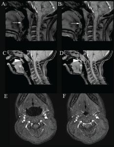
Figure 1. Cine magnetic resonance imaging showing upper airway obstruction. Sagittal images demonstrate the open (A, C) and narrowed airway (B, D) at the level of the tongue base (white arrow) and velum (arrow head)/tongue base (*). Posterior displacement of the tongue also caused posterior displacement of the soft palate (A, B). Axial images show the open (E) and narrowed airway (F) at the level of oropharynx/lateral walls (>) and tongue base (black arrow). Imaging technique prevents presentation of an example of concurrent obstruction at the level of the velum/oropharynx/lateral walls/tongue base in a single image.
Copyright 2017 The American Laryngological, Rhinological and Otological Society, Inc.
Cine MRI and DISE documented single and multiple sites of UAO in children with persistent OSA after AT. Cine MRI and DISE findings were similar in the majority of the children.
Background: Children with obstructive sleep apnea (OSA) may have multiple sites of upper airway obstruction (UAO). A wide variety of techniques has been used to evaluate UAO. Cine magnetic resonance imaging (MRI) and DISE have been increasingly used to evaluate UAO in children with OSA.
Study design: Retrospective chart review of medical records of children who underwent DISE and cine MRI.
Setting: Division of Pediatric Otolaryngology, Children’s Medical Center, Dallas.
Synopsis: Data pertaining to demographics, past medical history, body mass index, polysomnography, findings of DISE, and cine MRI were obtained. Fifteen children (11 boys, four girls; aged 7 to 18 years) were identified. Comorbid conditions were Down syndrome in nine patients, cerebral palsy in one, attention deficit hyperactivity disorder in two, and asthma in three. Severity of OSA was moderate in five, and severe in 10. DISE and cine MRI showed the same UAO site in 10 patients: a single site (tongue) in nine and multiple sites (tongue and oropharynx/lateral walls) in one (See Figure 1).
DISE showed additional UAO sites undetected by cine MRI in three patients. Cine MRI showed additional UAO sites undetected by DISE in one patient. DISE and cine MRI showed different sites of obstruction in one patient.
Citation: Clark C, Ulualp SO. Multimodality assessment of upper airway obstruction in children with persistent obstructive sleep apnea after adenotonsillectomy. Laryngoscope. 2017;127: 1224–1230.