Explore This Issue
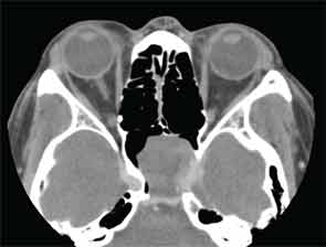
July 2013Presentation: A 53-year-old man presented with a one-month history of bilateral tinnitus. Magnetic resonance imaging (MRI) performed at another hospital detected a sphenoid tumor incidentally. The patient had no nasal symptoms, and endoscopic observation did not confirm a tumor in the nasal cavity. A contrast-enhanced computed tomography (CT) scan revealed a partially enhanced tumor in the left sphenoidal sinus that had destroyed part of the skull base (Figure 1).

Figure 1: CT scan showing partially enhanced tumor in left sphenoidal sinus.
The tumor also involved the internal carotid artery and contacted the optic nerve canal. T1/T2-weighted MRI showed a hypointense tumor, and a gadolinium-enhanced T1-weighted image showed a rounded mass. Although the patient experienced no visual abnormalities, a malignant tumor was suspected from the imaging findings.
—Submitted by Shuji Yokoyama, MD, Hiroshi Ogawa, MD, Takashi Matsuzuka, MD, Mika Nomoto, MD, and Koichi Omori, MD, department of otolaryngology, Fukushima Medical University School of Medicine, Fukushima, Japan.
What’s your diagnosis? How would you manage this patient? Go to the next page for discussion of this case.
Leave a Reply