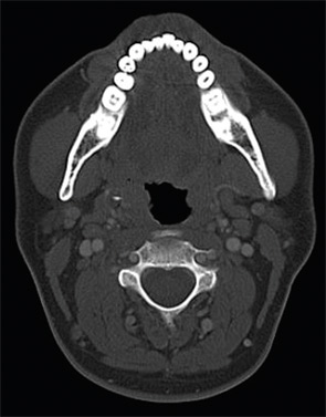
Figure 2: Axial CT scan.

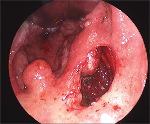
Figure 3: An arrow points to the styloid process.

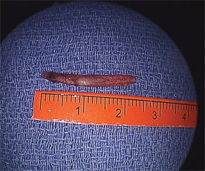
Figure 4: The styloid.
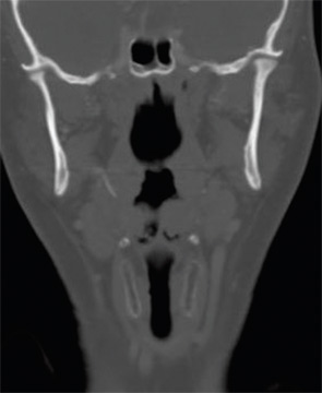
Presentation: A 33-year-old white male presented with a one-year history of right-sided odynophagia. Symptoms were constant and exacerbated by swallowing. He had a history of cryptic tonsils but had not undergone tonsillectomy; his past medical history was otherwise unremarkable. There was tenderness to palpation over the right tonsil with exacerbation of symptoms. No head and neck masses were appreciated. A CT scan was obtained (Figures 1, 2).
What’s your diagnosis? How would you manage this patient? Go to the next page for discussion of this case.

Leave a Reply