(click for larger image)
© Laryngoscope
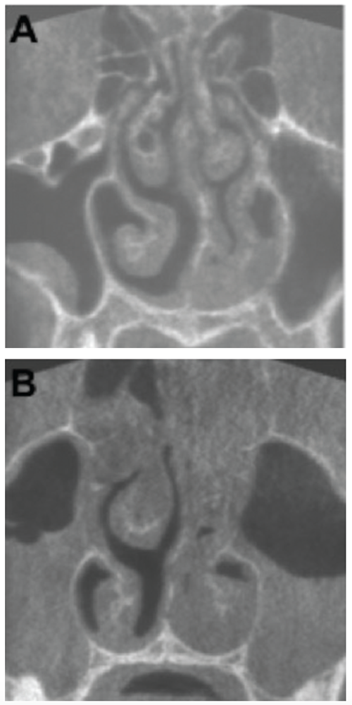
KA) Coronal plane of a CBCT scan from a patient three days after onset of symptoms. Haller cell and concha bullosa are present on the right side, and a cyst is present in the right maxillary sinus. The nasal septum is deviated to the left (19.4°). Both osteomeatal complexes are open, and other paranasal sinus mucosal abnormalities are not present. (B) Coronal plane of a CBCT scan from a patient nine days after onset of symptoms. There are air-liquid levels in the left maxillary sinus and gas bubbles in the right. The patient underwent maxillary sinus aspiration, which was cultured for Haemophilus influenzae.
What are the associations among paranasal imaging results, symptoms, bony anatomic variations, and culture-proven bacterial acute rhinosinusitis (ARS)?
Bottom Line: Paranasal mucosal abnormalities and osteomeatal complex (OMC) obstruction do not develop gradually during ARS but are present when symptoms begin and remain fairly constant in most patients both with and without bacterial ARS.
Explore This Issue
September 2016Background: ARS is a common condition, but its pathophysiology and the development of bacterial ARS are not yet completely known. Pathophysiology has earlier been evaluated with radiologic imaging, but systematic sequential imaging of the paranasal sinuses of the same patients during a single ARS episode would provide more information on ARS pathophysiology.
Study design: Inception cohort study with 50 conscripts with ARS diagnosed between February 1 and April 15, 2012.
Setting: Kajaani Garrison Health Centre, Kajaani, Finland.
Synopsis: Three sequential paranasal sinus scans using cone-beam computed tomography (CBCT) were performed during a single ARS episode for each patient. The median ARS symptom duration was two days at the time of the first CBCT scan, five days at the second, and 10 days at the third. Thirty-nine patients had respiratory virus nucleic acid in their nasopharynx at enrollment. Twenty patients underwent a maxillary sinus aspiration; in eight, the aspirate culture was positive for Haemophilus influenzae. At two to three days, 38% of patients had major abnormalities (most common in the maxillary sinuses); 42% had minor abnormalities in their paranasal sinuses. Sixty-eight percent had an occluded ostiomeatal complex (OMC) on at least one side. The total CBCT score had a weak correlation with the total nasal symptom score at nine to 10 days. Of individual symptoms, only the nasal blockage and clear discharge scores correlated with the total CBCT score. Smoking and a previous recurrent ARS history were not associated with total CBCT scores. At two to three days, patients with bacterial ARS had a slightly higher mean total CBCT score than those without. Limitations included unidentified viral etiology in some subjects, a relatively small study group, and a small-size scan field to reduce radiation dose.
Citation: Autio TJ, Koskenkorva T, Narkio M, et al. Imaging follow-up study of acute rhinosinusitis. Laryngoscope. 2016;126:1965-1970.