Explore This Issue
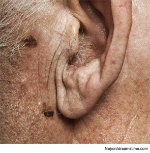
April 2012Presentation: A 75-year-old man presented with a left ear lobule melanoma and was found, on examination, to have a concurrent right parotid mass and bilateral cervical lymphadenopathy. A computed tomography (CT) scan of the neck confirmed a 2-cm right superficial parotid mass containing both solid and cystic components without any pathognomonic characteristics, as well as bilateral cervical lymphadenopathy with multiple 2- to 3-cm lymph nodes. Fine needle aspiration (FNA) biopsy of the parotid mass was consistent with carcinoma, whereas FNA samples from the cervical lymph nodes were non-diagnostic. The patient was taken to the operating room for excision of the left-ear melanoma, as well as a right superficial parotidectomy and right modified radical neck dissection. Intra-operative frozen pathologic analysis was performed on the parotid mass, level II lymph node and level IV lymph node. All three samples demonstrated poorly differentiated carcinoma as well as regions of monotonous lymphocytic proliferation.

The 75-year-old man presented with a left ear lobule melanoma.
—Submitted by Benjamin C. Paul, MD; Cameron L. Budenz, MD; Beverly Y. Wang, MD and David Myssiorek, MD. Drs. Paul and Myssiorek are with the department of otolaryngology, New York University; Dr. Budenz is with the department of otolaryngology, University of Michigan, Ann Arbor; and Dr. Wang is with the department of pathology, Beth Israel Medical Center, New York City.
What’s your diagnosis? How would you manage this patient? Go to the next page for discussion of this case.
Leave a Reply