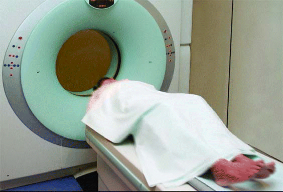
SAN DIEGO-With improved technology, as well as increased availability and access, diagnostic imaging has become the fastest growing segment of health care spending, with estimates of 15% to 35% increases annually. As a result, concerned third-party payers have established radiology guidelines, and some have teamed with radiology benefit management companies (RBMs) to bring down costs.
Explore This Issue
August 2007
In the field of otolaryngology, the most common imaging service used is computed tomography (CT), considered by many to be the gold standard diagnostic study for imaging of the paranasal sinuses for inflammatory disease.
According to Medicare databases, there’s been an increase of 61 percent in the use of CT studies from 2001 to 2005, said Brent Senior, MD, Associate Professor of Otolaryngology-Head and Neck Surgery at the University of North Carolina, Chapel Hill. In the same time period, otolaryngology usage increased 195 percent.
Dr. Senior’s remarks came during an April 27 panel presentation on CT scanning of the paranasal sinuses held during the American Rhinologic Society’s (ARS) program at the Combined Otolaryngology Spring Meetings.
Referring to radiology cost management, Dr. Senior noted that while this process of imaging service management is, in and of itself, not inherently flawed, the primary end point should not be cost savings, but improved patient outcomes. Cross-specialty cooperation in the development of evidence-based criteria for imaging services is essential to ensure that patients have access to proper diagnosis.
The usage patterns for CT in otolaryngology were the topic of panelist Pete Batra, MD, Assistant Professor of Surgery in the Section of Nasal and Sinus Disorders at the Head and Neck Institute of the Cleveland Clinic Foundation. Discussing the results of a Web-based, 20-question ARS survey, he noted that there is a wide variation in how we scan patients when they come into our offices.
In general, the survey showed a general trend of increased CT usage from pretreatment to post-first and second rounds of treatment. The most commonly used for diagnosis were 3-mm coronal (with and without axials), with plain films rarely used. MRI was utilized by only 32% of responders.
Additionally, the majority of physicians surveyed said they utilized hospital radiology or free-standing imaging centers for CT, with few indicating they had an in-office CT scanner or financial interest in an imaging center. Forty-six percent who own an in-office CT scanner reported reading, interpreting, and billing for the scans.
Leave a Reply